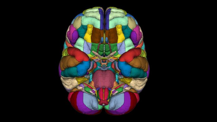
Scientists have created a detailed cellular atlas of the basal ganglia, a group of deep brain structures involved in movement, decision-making, emotions and habit formation. The work provides researchers with a reference map of the cell types in this region, the genes active within them and the DNA regulatory switches that control that activity.
The research, presented in more than two dozen studies published in Cell and related journals, was conducted as part of the US National Institutes of Health-supported BRAIN Initiative. It is described as the 1st comprehensive cross-species cellular roadmap for any brain region.
What the basal ganglia do
The basal ganglia are an interconnected set of structures deep within the brain. They include the striatum, globus pallidus and substantia nigra.
Together, these structures help select and start movements. They also support habit formation, reward learning, motivation and emotion.
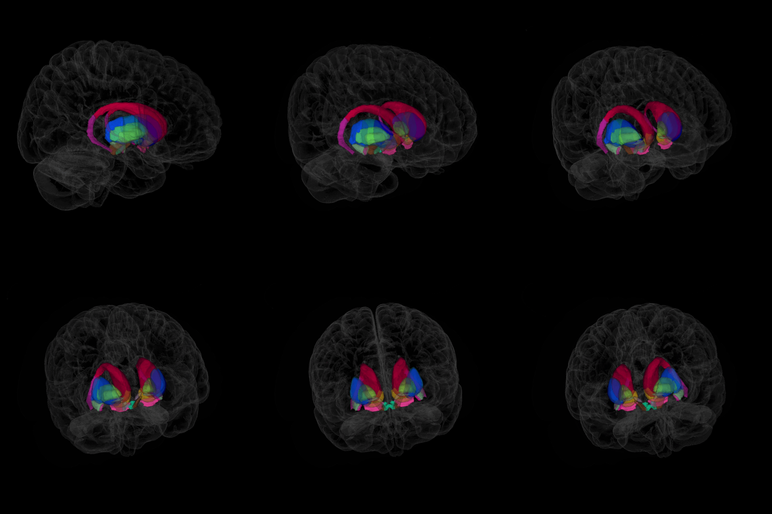
Dysfunction of the basal ganglia is associated with several major conditions, including Parkinson’s disease, Huntington’s disease, schizophrenia, Tourette’s syndrome, bipolar disorder, obsessive-compulsive disorder and addiction.
What scientists mapped
The researchers mapped cell types and cellular gene activity in the basal ganglia of humans, mice, macaques and marmosets. Using brain tissue from deceased human donors, they examined 21 million cells in the human basal ganglia, along with millions more cells in the other three species.
The atlas brings together several layers of information:
Gene activity – Which genes are switched on in individual cells
Epigenome – Chemical compounds and proteins attached to DNA that regulate gene activity
Spatial maps – Where each cell type is located within basal ganglia tissue
Why cross-species comparison matters
Much of what was previously known about the basal ganglia, and much early testing of new treatments, came from research in mice and monkeys. Many studies cannot ethically be performed in living human brains.
By mapping the same brain region across humans, mice, macaques and marmosets, researchers can compare cellular composition, function, connectivity patterns and responses to pharmacological interventions across species.
What the atlas may reveal
The researchers identified a new type of brain cell associated with schizophrenia and bipolar disorder. They also documented genetic changes in specific cell types that could play a role in autism and found changes in gene activity in certain cells associated with ageing.
Most DNA variants linked to brain disorders lie outside the protein-coding parts of genes. Many are located in DNA “switches” that control when and where genes turn on, and those switches are often active only in particular cell types.
An atlas of this kind can help researchers trace a disease-risk DNA variant to the specific nerve cell or support cell in which it acts. According to Joseph Ecker, a molecular biologist and geneticist at the Salk Institute, that is a first step toward understanding how a variant contributes to disease and, eventually, toward treating it.
Important limitations
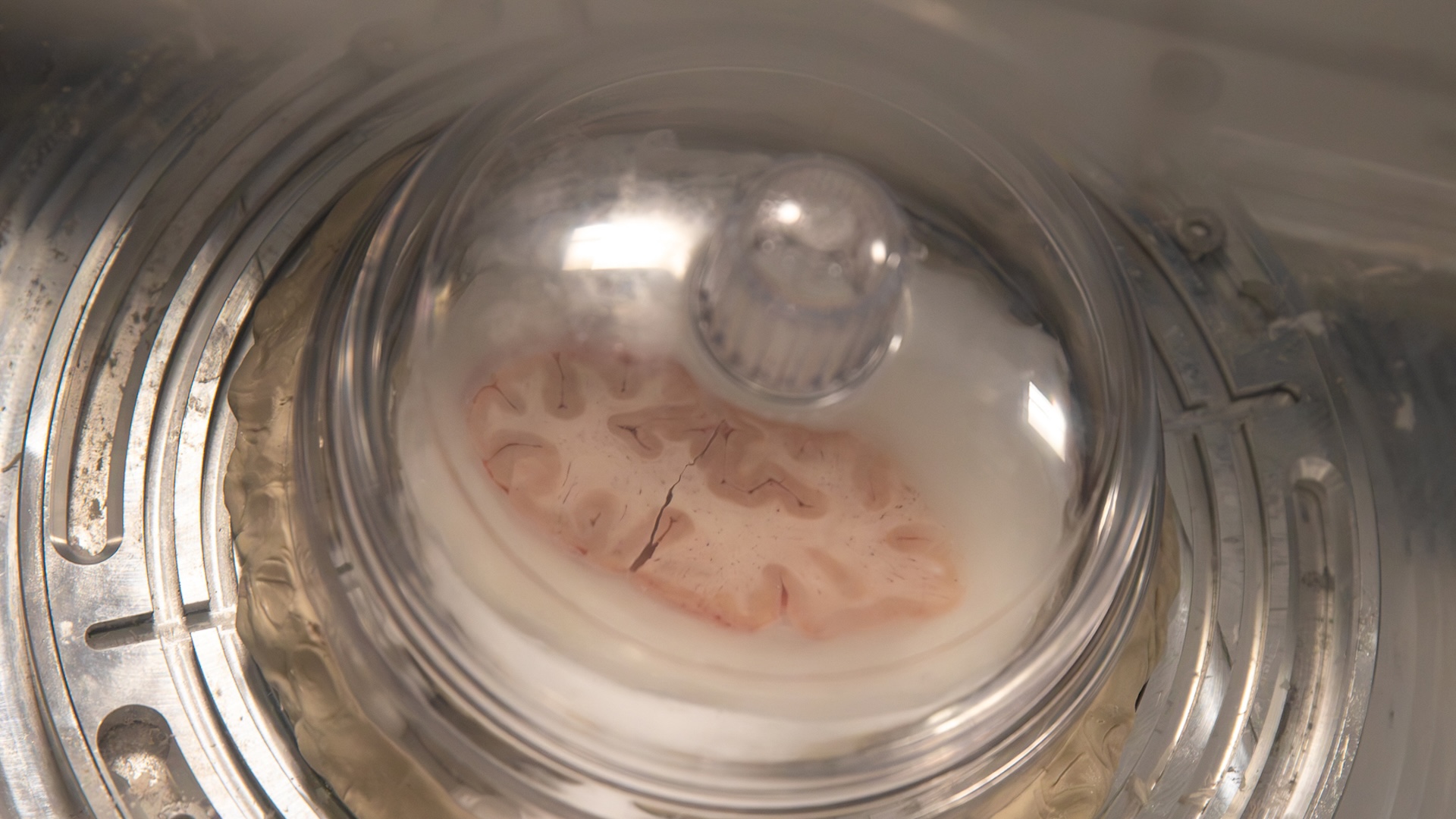
The researchers emphasise that the atlas is a foundation, not a finished product. Many datasets come from postmortem tissue, where donor history, tissue quality, agonal state, preservation method and sampling strategy can influence molecular profiles and complicate comparisons between donors.
Coverage is also uneven across species, developmental stages, anatomical regions and data types. Although transcriptomic, epigenomic, spatial and electrophysiological datasets are increasingly integrated, each has different resolution, sensitivity, sparsity and technical biases. Aligning these datasets remains a major computational and conceptual challenge.
Cross-species comparisons are powerful but imperfect. Homologous cell types may differ between species in abundance, spatial distribution, regulatory logic and functional role.
The researchers also note that the atlas should be viewed as a living reference system rather than a static product. It will need continued refinement through broader sampling, functional validation, integration with disease data, transparent versioning and community contribution.
In the longer term, such a map could help narrow the search for disease mechanisms and potential therapeutic targets. But the researchers are clear that translating these findings into safe and effective treatments will take years.
Source: Cell; Cell; Allen Institute