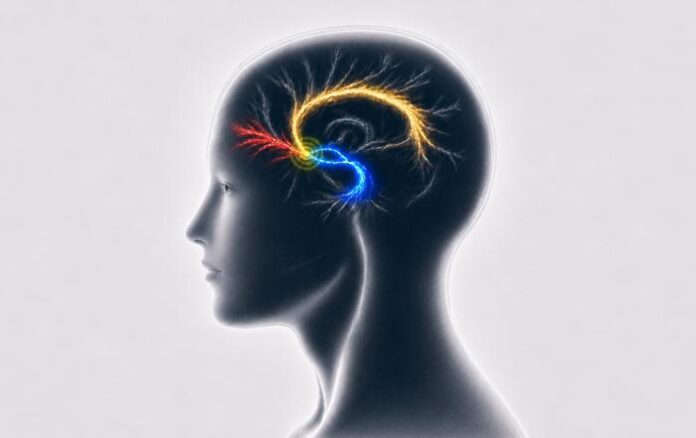
Scientists at Mount Sinai in New York have identified three white matter fiber tracts in the brain whose stimulation is associated with sustained clinical improvement in patients with treatment-resistant depression.
The discovery is based on deep brain stimulation (DBS), an experimental method in which electrodes are surgically implanted in the brain to deliver electrical pulses to neural tissue. The goal is to target specific brain circuits.
While deep brain stimulation is associated with long-term symptom improvement in some patients with treatment-resistant depression, clinical outcomes vary, and symptoms often fail to improve adequately despite standard care.
A central challenge in using deep brain stimulation is determining which brain pathways need to be stimulated to achieve the desired outcome. The electrical field affects more than just the tissue immediately surrounding the electrode; its impact extends to whichever nerve fibers fall within the stimulation field.
Three Brain Pathways
Researchers analyzed data from 131 patients across three independent cohorts who had previously undergone subcallosal cingulate (SCC) deep brain stimulation. Located on the inner surface of the cerebral hemispheres directly beneath the fronto-inferior part of the corpus callosum, this region belongs to the limbic system and plays a key role in regulating emotions, mood, and behavioral responses to stress.
By combining models of electrode-generated electrical fields with brain connectivity maps, the researchers identified three white matter pathways whose stimulation was linked to sustained clinical improvement:
The cingulum bundle connects various brain regions, including limbic (emotional) structures and cortical areas, and is involved in regulating emotion, attention, motivation, and mood;
The forceps minor connects corresponding areas of the right and left frontal lobes, facilitating information exchange between them. These connections participate in cognitive processes, including the regulation of thought and behavior;
The medial uncinate fasciculus and ventral amygdalofugal pathway are neural pathways involved in connecting the amygdala with regions of the frontal lobe, thalamus, and hypothalamus. These connections are engaged in processing emotions, including fear and stress-related responses.
How Does Stimulation Affect Brain Pathways?
Brain white matter consists primarily of bundles of long nerve cell projections (axons). Most of these fibers are covered by a myelin sheath that facilitates rapid transmission of nerve impulses. They link different brain regions together, while the structural connectome serves as a map of these anatomical connections.
To assess treatment efficacy, researchers relied on clinical response, defined as at least a 50% reduction in symptoms measured on a standard depression rating scale (HAM-D17).
The study showed that patients whose electrical stimulation covered all three identified pathways experienced sustained clinical improvement during long-term follow-up.
Additionally, a significant difference emerged between treatment responders and non-responders: in patients for whom treatment was not successful, electrical stimulation insufficiently engaged the left cingulum bundle and medial uncinate fasciculus.
This finding explains why treatment outcomes can differ even when electrodes are placed in similar anatomical regions: alongside electrode location, whether the generated electrical field reaches the necessary nerve fibers is critical.
A Personalized Treatment Approach
The results support using individual structural brain connectivity maps during treatment planning rather than relying solely on general anatomical landmarks, allowing electrode placement to target intended neural pathways more precisely.
Study Limitations and Future Prospects
Importantly, deep brain stimulation remains an experimental approach for treating depression and is not an approved standard treatment.
The study did not test whether repositioning electrodes would improve outcomes for patients whose stimulation fell short of the necessary pathways. Consequently, the findings point to an observed link between pathway stimulation and clinical improvement, but do not prove that engaging these pathways alone is sufficient—other factors may also influence treatment response.
The study also had a limitation: individual diffusion MRI brain scans were unavailable for some patients, requiring researchers to rely on generalized structural connectivity maps for part of the analysis. However, among patients with individual scanning data available, using individual versus generalized maps yielded similar results.
Overall, the findings demonstrate that stimulating specific brain pathways is essential for treatment efficacy. Future studies should evaluate how data on individual structural and functional brain connections can refine patient selection, electrode placement, and stimulation parameter settings.