


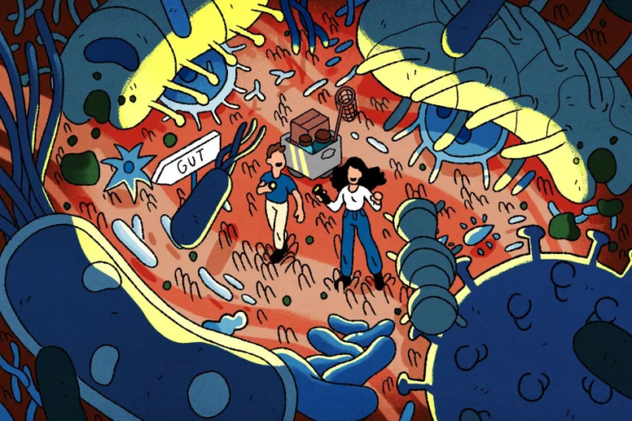
Traditionally, the muscular layer of the gut (muscularis) was viewed merely as the organ’s “mechanical engine,” whose sole function was the peristaltic contractions required to propel food forward. Modern science, however, now considers this view outdated.
Today, the muscularis emerges as a far more complex, multicellular regulatory system. It is a dynamic environment where smooth muscle cells, enteric neurons, glial and immune cells, and microbial signals remain in constant communication. It is precisely their coordinated interaction that determines gut motility, tissue homeostasis, and symptom generation.
This conceptual shift is fundamental: it no longer views intestinal pathologies as isolated defects of muscle or nerve, but rather frames them as systemic “communication breakdowns” occurring at the tissue level.
The Dynamic Ecosystem of the Gut Wall
The muscularis externa is an integrated neuroimmune-muscular system where local signals are continuously processed and converted into precise, coordinated physiological responses.
This approach makes it clear how the exact same tissue manages to ensure normal peristalsis in a healthy organism, trigger pain and hypersensitivity in irritable bowel syndrome (IBS), engage in fibrotic remodeling during inflammatory bowel diseases (IBD), and suffer functional disruption following infection or surgical intervention. This demonstrates that the muscularis is not merely an effector—it is an intrinsic part of the control system itself.
Parallel to this shift, research methods are being radically refined. The latest single-cell and spatial analyses have shown that the muscularis is by no means a uniform tissue; rather, it unites numerous small, unique cellular microenvironments.
Discoveries like these have allowed science to replace the vague, general diagnoses of “motility disorders” from the past with clear cellular-level precision. In practice, this means one thing: the medicine and therapeutics of the future will become strictly personalized, taking into account exactly which cellular types, signaling pathways, or microniches are disrupted in each individual patient.
Cellular Architecture
The muscular layer of the gut is not as simple in structure as it might seem at first glance: it represents a complex, tightly intertwined network of smooth muscle cells, neurons, support cells, and immune agents. Every participant in this system has a strictly defined role—neurons control muscle contraction and relaxation, specialized cells (interstitial cells of Cajal) dictate their functional rhythm, and supporting elements care for neuronal behavior and survival.
In this context, a deeper understanding of the role of muscularis macrophages is particularly interesting. They are not merely passive observers of inflammatory processes; they play a decisive role in neuronal survival and the preservation of motility. Furthermore, depending on the developmental stage or inflammatory context, they can radically alter their functional profile and adapt their role to the situation.
This specialization of macrophages is one of the most vivid examples of how knowledge in this field has deepened. These cells resemble other resident tissue macrophages in that they are long-lived and locally programmed, yet they possess a remarkable capacity to support the enteric nervous system. During development, they participate in establishing neuronal circuits, while in adulthood, they contribute to their maintenance. Thus, the concept that the muscularis contains its own intrinsic system for preserving neuromuscular integrity gains further ground.
This holds clear practical significance for clinical medicine. In disorders such as diabetic gastroparesis, postoperative ileus, and inflammatory bowel disease, the loss of protective macrophage phenotypes or improper macrophage activation can contribute to motility impairment and neuronal damage. This implies that these pathologies can no longer be viewed solely through the lens of smooth muscle dysfunction; they may instead result from a breakdown in the local support system that normally safeguards the neuromuscular apparatus. This represents a fundamental shift in perspective, expanding the scope of potential therapeutic targets.
The Neuro-Immune Connection
Equally fascinating is the bidirectional connection between enteric neurons and immune cells. Neurons can determine the composition and behavior of the immune system, while immune mediators can alter neuronal excitability, glial activity, and smooth muscle function. Consequently, we are dealing with a reciprocal system in which immune and neural signals continuously balance one another. This helps explain why intestinal diseases are frequently accompanied by simultaneous changes in motility, inflammatory processes, and sensitivity, rather than a single, isolated impairment.
It is rightly emphasized that different subsets of neurons can exert distinct influences on immune cells. This nuance adds greater precision to earlier works that described “neuroimmune interactions” only in general terms. It also suggests that disease may develop when an improper signal is activated in the wrong cell or tissue layer.
What This Changes in Practice
Theoretical discoveries are only the beginning—what matters most is how they translate into real medical practice and patient management.
In Crohn’s disease, the inflammatory process extends beyond the mucosa. Inflammation reaches deep into the muscular layer, causing inflammation of the neural plexuses (plexitis), neuronal damage, glial network activation, muscle remodeling, and fibrosis. A decisive role in this process is played by so-called “creeping fat,” which clearly demonstrates that mesenteric adipose tissue actively participates in the development of fibrosis and strictures. This explains the clinical paradox of why mechanical obstruction develops in patients even when mucosal inflammation appears to be under control.
In ulcerative colitis, the picture appears different at first glance, as lesions classically involve only the mucosa. However, research clearly shows that “waves” of chronic inflammation and oxidative stress extend into deeper layers, disrupting the function of the muscularis as well. This confirms once again that strict clinical categories (“mucosa only” vs. “muscle only”) often do not reflect the true biological reality. This is why, in practice, a patient may experience persistent symptoms and functional impairments even alongside a visually clear mucosa on endoscopy.
Irritable Bowel Syndrome (IBS)
Today, Irritable Bowel Syndrome is recognized as a disorder of gut-brain interaction, rather than merely a motility issue or a “functional” condition in the outdated sense. The muscularis occupies a central place in this new understanding, as it serves as the convergence point for disrupted gut-brain signaling, immune activation, epithelial anomalies, microbiome shifts, and biological stress factors, which ultimately lead to pathological muscle contractions and visceral hypersensitivity.
This model also accounts for the heterogeneity of IBS. In some patients, smooth muscle hyperexcitability may predominate, while in others, sensory hypersensitivity, serotonergic dysregulation, mast cell activity, or microbiome-derived signals may be more significant.
The practical value of such an approach is that it facilitates more accurate phenotyping and a more rational selection of treatment. Furthermore, it helps clinicians take patient symptoms seriously rather than reducing them strictly to psychogenic factors or simple motility disturbances.
Infection and Reconstruction
Discussing infections adds an entirely new, compelling dimension to the subject:
Helminthic (parasitic) infections hyper-excite muscle tissue and induce a hypercontractile response (exaggerated, forceful intestinal contractions) aimed at expelling the parasite.
Viral infections, together with the immune system, act more directly toward damaging neurons.
Bacterial infections may result in severe tissue damage or, conversely, transition into a state of tolerance.
This diversity is critical because it indicates that post-infectious motility disorders (post-infectious dysmotility) do not represent a single, uniform pathology, but rather a group of disorders with distinct cellular origins.
Pathologies like Hirschsprung’s disease display a similar level of complexity and have long expanded beyond classic concepts where the problem was attributed solely to the absence of nerve ganglia (aganglionosis). Research clearly shows that the pathology encompasses fibrosis, stromal reconstruction, glial alterations, and impaired epithelial regeneration.
This holds immense clinical significance, as it clearly demonstrates that surgical resection of the aganglionic segment does not automatically eliminate all tissue anomalies. This is precisely why the idea of restoring the enteric nervous system through cell-based therapy warrants special attention, shifting the discourse from strictly surgical intervention toward regenerative therapeutics.
Smooth Muscle and the Microbiota
Smooth muscle cells do not function solely by contracting in response to nerve impulses; they also participate in tissue repair and respond instantaneously to immune signals. In turn, immune cells themselves possess the capacity to drive muscle contraction directly, sometimes even without nervous system involvement. A striking example of this is specific receptor (TRPV4) signaling in macrophages, which directly triggers prostaglandin release and induces direct smooth muscle contraction.
However, behind this cellular dialogue stands another powerful player. Microbial metabolites—specifically short-chain fatty acids and tryptophan derivatives—directly determine neuronal maturation, smooth muscle excitability, and macrophage phenotype. This turns the gut microbiota into an active regulator of muscularis function rather than a passive bystander. In disorders such as IBS and post-infectious dysmotility, this approach validates the logic of microbiome-targeted strategies, albeit in a far more cautious, scientifically grounded manner than a simple enthusiasm for probiotics.
Against this backdrop, future progress will likely be achieved not by targeting individual cells in isolation, but by managing communication networks. This could involve reprogramming macrophage phenotypes, modulating neuropeptidergic pathways, restoring the balance of microbial metabolites, or identifying tissue-specific biomarkers that distinguish one muscularis pathology from another.
This approach also indicates that therapy must align with the dominant biological process characteristic of an individual patient, rather than being applied as though all motility disorders share the exact same underlying mechanism.
Current research bridges molecular mechanisms and clinical practice without exaggerating the field’s present state of advancement. Most of the strongest evidence still relies on animal models, and the validation of these mechanisms within the human body remains incomplete. Nevertheless, the trajectory of development is clear: the muscularis is establishing itself as a tissue where imaging, transcriptomics, microbiology, immunology, and physiology may finally unite to implement more precise diagnostics and therapeutics.
Source: nature
