


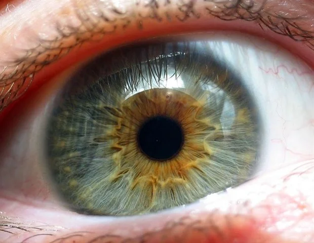
The formation of the human visual system is one of the most refined and precise processes. Its functional perfection is directly dependent on how effectively the organism manages, during the period of embryonic development, to create that filigree configuration of cells that determines our “super-vision.” This process relies on an exact sequence of molecular signals, which ultimately determines the structural formation of the retina’s most sensitive point, the foveola.
Precisely this microscopic area determines our ability to perceive the world in high resolution. The foveola is a small depression in the central part of the retina, the macula, which allows us to see the smallest details and accurately recognize facial expressions. In the foveola, M/L type cones (green and red color-perceiving cells) are arranged with extremely high density, while “blue” (S) cones, rods, and blood vessels are practically not found in this area.
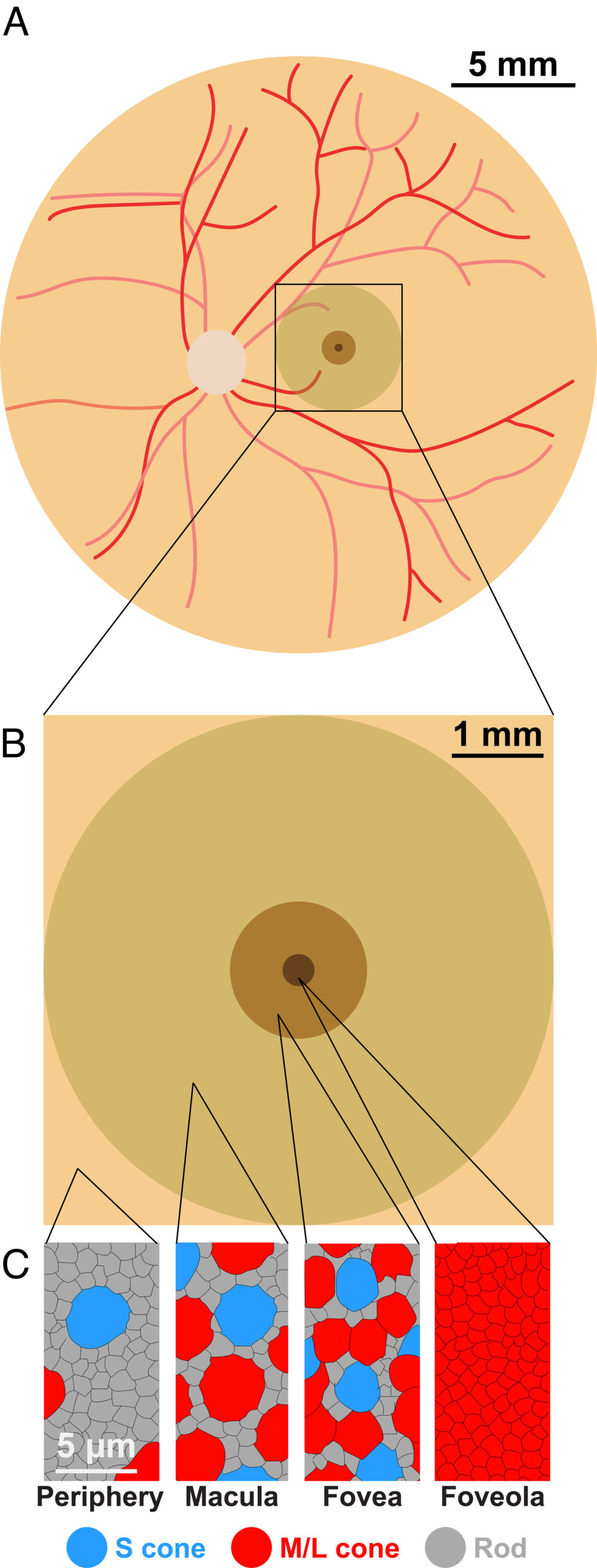
A new study explains the process of forming this unique architecture. Scientists were able to identify the molecular signals that determine the specialization of visual cells and their functional differentiation during the embryonic period.
The Secret of Cellular Transformation
According to the traditional view, the specialization of retinal cells was considered a linear and irreversible process. Scientists believed that a strict division occurred at the very initial stage of development: a cell received either an S-type (blue) or an M/L-type (green/red) identity. According to this model, once a cell chose the “blue” trajectory, its transformation into another type of receptor was impossible.
However, cellular analysis of the fetal retina called this opinion into question. It turned out that at the 11th week of embryonic development, S-cones appear en masse in the embryonic area of the future foveola. These data are in clear discrepancy with the architecture of the adult human retina, whose central area is completely free of “blue” cones.
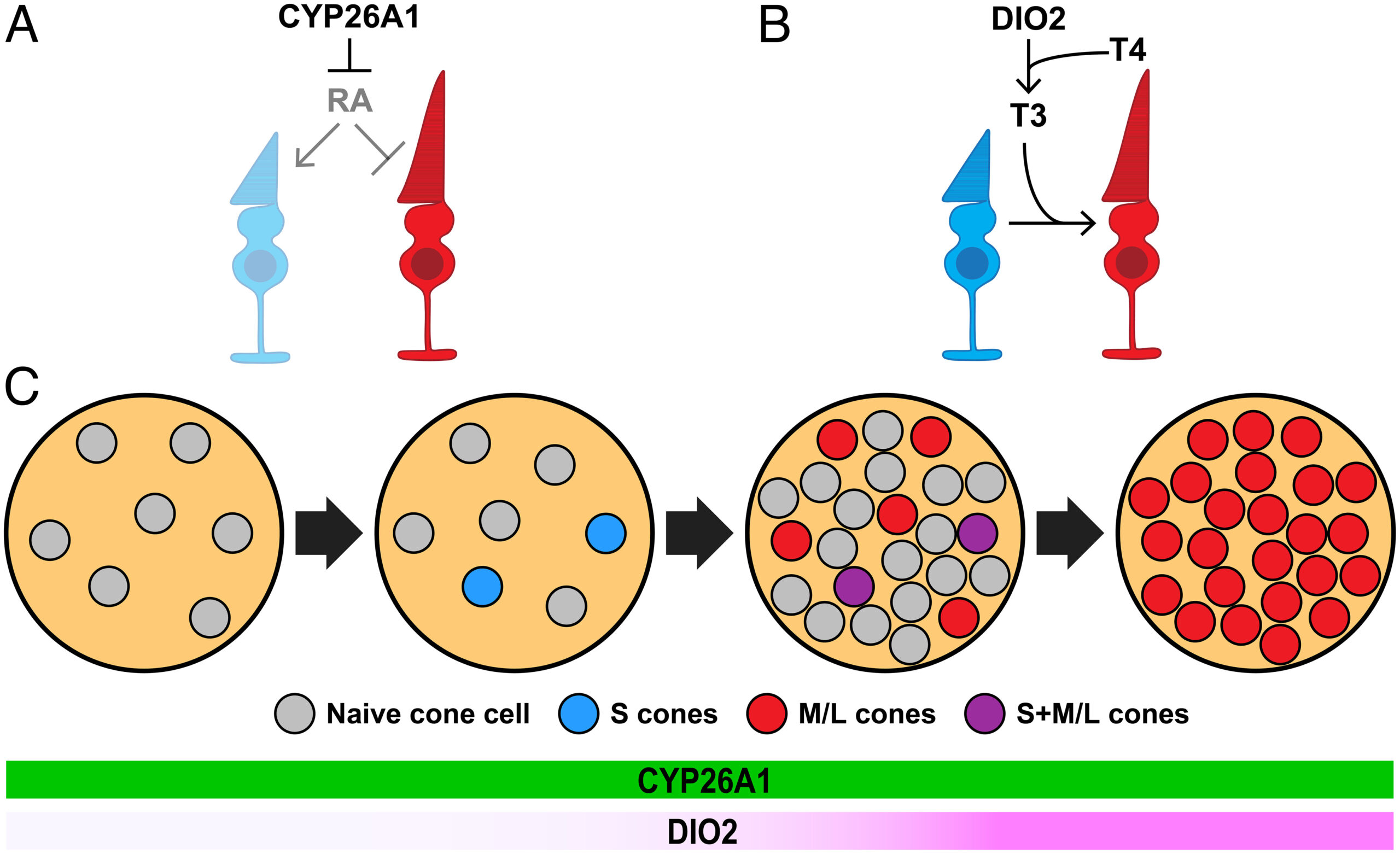
This paradoxical given gave rise to a logical question: by what mechanism do the mentioned cells disappear from the central area? An early hypothesis explained this process through physical migration, and it was believed that the S-cones moved toward the periphery. However, the latest data revealed a unique case of cellular plasticity. It turned out that instead of moving, the photoreceptors undergo molecular reprogramming and finally form into M/L cones.
Photoreceptor Dynamics
Molecular research of the fetal retina revealed three different populations of cones: S-type, M/L-type, and so-called co-expressive cells, which simultaneously produce both types of opsin. Precisely the latter represents the transitional link in the process of cellular transformation.
The chronology of development shows that at the 10th week, the first S-cones appear in the embryonic area of the foveola, although by the 14th week the picture changes radically: differentiated M/L cones occupy the dominant position in the center of the foveola, while a high concentration of co-expressive cells is noted next to them.
At the completion of the process, at the stage of adulthood, the central zone of the foveola is already exclusively composed of M/L cones. Taking into account that no sign of cell death (apoptosis) was revealed until the 23rd week of development, scientists concluded that the early S-cones do not “die” or physically move, but rather change their differentiation trajectory and transform into M/L type receptors.
Retinoic Acid and Hormonal Regulation
The formation of the foveola’s unique structure depends on two main factors that coordinately manage the differentiation of photoreceptors. The first of these is the local degradation of retinoic acid (RA), which is carried out by the enzyme CYP26A1. The high activity of this enzyme in the central retina causes the intensive breakdown of retinoic acid, which is critically important for limiting the initial formation of “blue” cones.
Experimental models confirmed that a high concentration of retinoic acid stimulates the development of S-type receptors, while its deficiency, which is artificially created in the foveola area, opens the way for the dominance of M/L type cells.
The second decisive link in the process is the thyroid hormone (TH), specifically its active form—T3. It turned out that this hormone suppresses the genes expressing S-identity and facilitates the transformation of cells into M/L type receptors. The long-term impact of hormonal signals “locks” the cells in their final state, which ensures the functional stability of the foveola. Thanks to this molecular “engineering,” the organism manages to create a cellular configuration in the center of the retina that is responsible for the exceptional sharpness of our vision.
Therapeutic Perspective
By using retinal organoids, “mini-organs” cultivated in laboratory conditions, scientists were able to achieve a full molecular imitation of the foveola formation process. This achievement fundamentally changes our approach to the treatment of ophthalmological diseases. Specifically, deciphering molecular mechanisms opens the way for targeted hormonal therapy, which is critically important for the prevention of visual pathologies in premature newborns.
The knowledge obtained directly reflects on the quality of retinal organoids, which makes them ideal material for future transplantation. The mentioned breakthrough lays the foundation for personalized medicine, where correcting damage caused by macular degeneration already appears as a real scientific perspective.
Source: PNAS
