


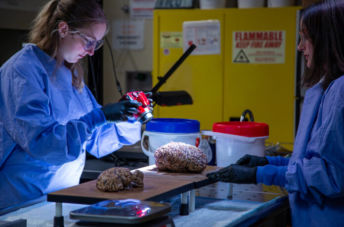
The Allen Institute is launching a large-scale brain mapping project designed to detect neurodegenerative processes at their inception and accelerate the development of novel therapeutics. This ambitious 14-year initiative, backed by $200 million in funding, will significantly enrich the organization’s existing foundation, the Seattle Alzheimer’s Disease Brain Cell Atlas (SEA-AD). Within the framework of the project, researchers will examine the postmortem brain tissue of thousands of donors diagnosed with Parkinson’s disease, Huntington’s disease, and amyotrophic lateral sclerosis (ALS).
Historically, in the study of neurodegenerative diseases, scientists have focused primarily on pathological protein aggregates—such as amyloid and tau in Alzheimer’s disease, and alpha-synuclein in Parkinson’s disease. The Allen Institute, however, is approaching the dilemma from a fundamentally different perspective. They are investigating changes occurring at the level of specific neural circuits and target cells during the earliest stages of the disease, long before toxic protein deposits manifest.
Dr. Ed Lein, the project lead, is confident that such granular mapping of cellular architecture will unveil novel therapeutic targets and identify universal pathways to treatment capable of addressing multiple disorders simultaneously.
This initiative builds upon the successful precedent of the SEA-AD program, yet with a doubled budget, it encompasses a significantly broader spectrum of pathologies. Investigators will perform comparative cellular and molecular analyses on tissues from both healthy and diseased donors. The massive datasets generated through this process will be processed by artificial intelligence, facilitating the early detection of structural damage in the most vulnerable cellular subpopulations.
The Translational Bridge
A core advantage of the project lies in the parallel examination and cross-species comparison of primate and rodent brain architectures. Forging these interspecies parallels will help clarify precisely which human cell types are recapitulated in animal models, thereby substantially reducing ambiguity during preclinical drug testing. Scientists anticipate that these baseline reference data will drastically accelerate laboratory research. Optimally, this could enable the first human clinical trials of novel therapeutic strategies within just a few years.
The brain health accelerator unites 28 partner institutions—including universities, non-profit organizations, and scientific research centers. This coalition operates under uniform, centralized standards for tissue procurement, biospecimen banking, and biodata sharing. Partners will undergo specialized training to standardize workflows for handling postmortem donor tissues, enabling high-fidelity RNA sequencing and other high-precision assays. Furthermore, the program integrates clinical histories and neuroimaging data. This comprehensive synthesis will allow researchers to map correlations between the brain’s cellular characteristics and the patient’s real-world clinical symptoms.
The scientific community has expressed strong optimism regarding the scale and transformative potential of this initiative. Leadership at the NIH BRAIN Initiative emphasizes that detailed cellular atlases are indispensable for deciphering how and why brain cells cease to function normally. Clinicians specializing in frontotemporal dementia and other neurodegenerative disorders hope that these cellular and network roadmaps will illuminate novel, personalized therapeutic modalities.
Nevertheless, investigators face formidable practical hurdles. Procuring large volumes of postmortem tissue accompanied by comprehensive medical histories and neuroimaging records demands immense logistical coordination. Standardizing biospecimen preservation across disparate research centers, ensuring demographically representative cohorts, and navigating legal constraints surrounding data privacy and sharing present additional challenges. Ultimately, translating purely descriptive atlases into actionable medical targets will require rigorous, hypothesis-driven validation in both laboratory models and human tissues.
Source: Science
