


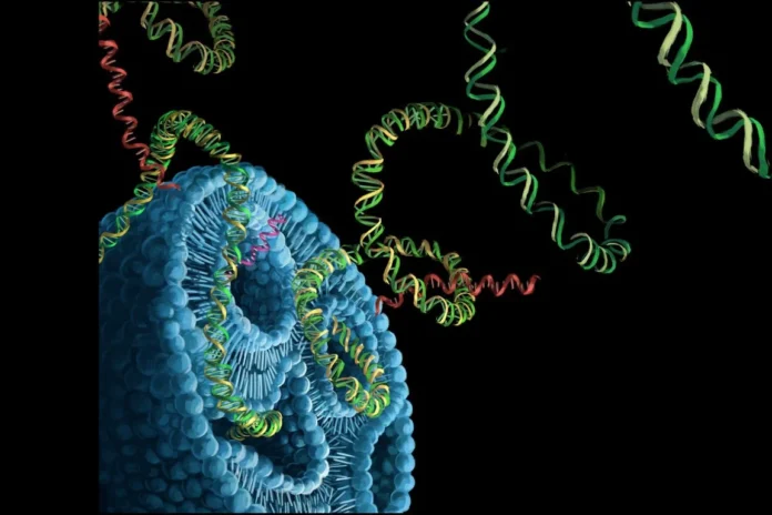
In modern medicine, determining the risk of breast cancer relies primarily on statistical data and the visualization of already formed pathological foci. For example, mammographic screening maintains its effectiveness only when a cancerous growth physically exists. At the same time, genetic testing provides a real answer for only a small portion of patients, approximately 5-10%. As a result, the vast majority of women, who do not exhibit distinct hereditary factors or radiological changes, remain facing a practically “invisible” threat.
To eliminate this existing diagnostic gap, scientists from City of Hope and Berkeley shifted their attention to the biophysical characteristics of the cell. They hypothesized that before visual malignant transformation begins, the cell first undergoes structural changes and loses its elasticity, which the researchers have termed “mechanical age.”
The practical evaluation of this theoretical phenomenon occurs through the MechanoAge platform, using a kind of “cellular stress test.” Inside the device, each cell placed in a microfluidic chip passes through a special narrow channel, during which artificial intelligence observes its speed of deformation and its elasticity.
During this process, the system records the most important parameter – the time required for the cell to regain its original shape. It is this data that becomes an indicator of how far the structural aging of the cell has progressed and, consequently, how high the risk of developing cancer is.
Designing the Mechano-NPS Platform
The foundation of the study is the methodology of mechano-node-pore sensing (mechano-NPS). The primary instrument of the process is a microfluidic chip, which externally resembles a small glass plate. Integrated into its internal space are channels much thinner than a human hair, equipped with platinum and gold electrodes. This technology allows scientists to precisely record any movement and structural transformation of the cell along with its volume.
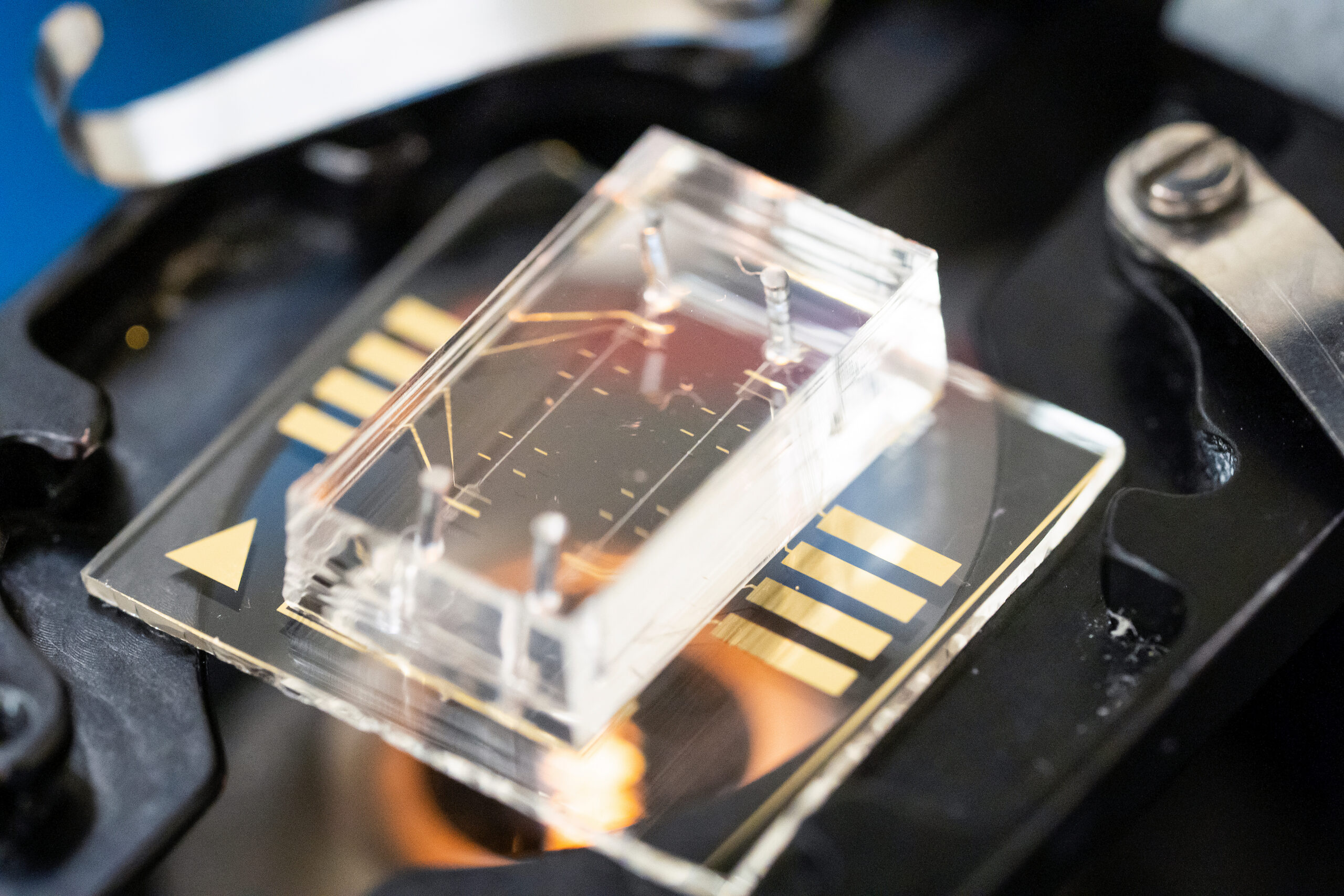
The uniqueness of this method lies in the fact that it does not cause damage to the cell. The mechanical force used is only sufficient to change its shape, which does not threaten the integrity of the structure. Ultimately, the researchers obtain a complete picture of the data and reveal those hidden threats that often remain unnoticed when using traditional visual methods.
How Does the Apparatus “See” the Cell?
To measure the elasticity of cells, the system uses a 1-volt direct current, which produces specific electrical signals as the cell passes through the channel. A specialized program instantaneously deciphers these impulses and determines several key parameters: the size of the cell, its degree of compression, and the time required for shape recovery. In order for the data to be objective and to ensure that cell size does not have a misleading influence on the result, a special dimensionless metric of deformability is used. This indicator allows scientists to accurately calculate the stiffness of the cell and to equalize the data so that the comparison of cells of different sizes occurs reliably.
MechanoAge: From Data Analysis to Precise Prediction
The process begins with the preparation of “learning material,” for which the researchers collected nearly 2,000 data units based on cells obtained from 18 donors.
In the first stage of information processing, each cell was assigned a corresponding age mark. This stage was necessary so that the computer system could learn to distinguish the visual or structural characteristics of healthy and damaged cells of a specific age.
In the next step, to prevent statistical errors, the researchers carried out “cleaning” of the data using the Yeo-Johnson transformation and downsampling methods. This approach involves a systemic equalization of information during which no subgroup—for example, young cells—is assigned a quantitative advantage. It is this balancing of the database that ensures that the final analysis is as objective and reliable as possible.
To achieve maximum accuracy, the scientists chose not one specific program, but a GBM meta-model, which unites three different types of algorithms. This approach is known as an “ensemble” model and, in essence, resembles a joint evaluation of a patient’s condition by several doctors and the bringing forth of a single, specified conclusion.
After completing the formation of the model, its rigorous checking stage began, which was carried out using the 10-fold cross-validation method. This process involves testing the program ten times on various combinations of data to confirm the reliability of the system’s operation.
The Role of the KRT14 Protein in Cellular Aging
The protein keratin 14 (KRT14) plays a decisive role in the aging process of the cytoskeleton. Scientists noticed that as age increases, this protein accumulates excessively in luminal cells. To prove that KRT14 itself is the cause of “aging” and not a mere accompanying phenomenon, the researchers artificially altered its levels.
Experimental manipulations confirmed that an increase in keratin levels in young cells determines their mechanical aging and an increase in the risk (RISQ) indicator. Conversely, the suppression of this protein in older cells caused a kind of “rejuvenation” effect—the share of “aged” cells in the database decreased from 85% to 15-23%.
Future Plans
When interpreting the results obtained, the methodological framework of the study and its limits must be taken into account. Specifically, the small volume of the research cohort and limited demographic diversity raise the need for verification of the data in larger populations. Furthermore, the ex vivo format of the experiments indicates that indicators obtained in laboratory conditions may not fully correspond to the physiological processes ongoing in the organism.
However, these limitations represent strategic orientations for the scientists for the further development of the project. The next stages of research aim to adapt the method to less invasive procedures, such as fine-needle aspiration (FNA) biopsy, which will make the technology more clinically accessible. Future work also includes the integration of multiplex technologies and the in-depth study of various ethnic groups, which will finally confirm the universality and diagnostic accuracy of the Mechano-RISQ metric.
Full study available: Lancet eBioMedicine
