


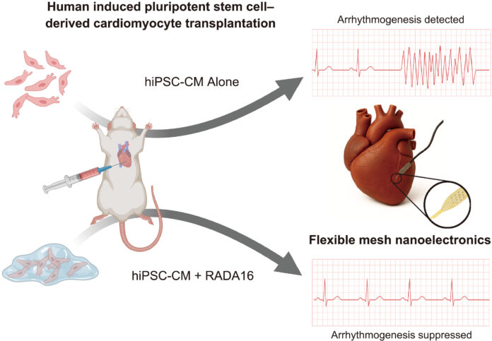
A team of researchers at Harvard University has created an innovative platform that has made it possible to overcome one of the main challenges in repairing damaged heart tissue: the synchronization of transplanted stem cells with the patient’s native heart cells.
For decades, heart muscle cells grown from a patient’s stem cells (hiPSC-CMs) have been considered a promising approach for regenerating the organ following a heart attack or heart failure. However, the transplantation of these cells was often accompanied by a risk of arrhythmia, as the new cells failed to properly integrate with the rhythm of the host tissue.
As part of a joint study between Harvard’s School of Engineering and Applied Sciences (SEAS) and the Department of Stem Cell and Regenerative Biology, researchers, led by Professor Jia Liu, developed the first platform that enables continuous monitoring of the maturation, communication, and synchronization of cells transplanted in vivo (in a living organism).
The researchers used specialized “high-tech mini-organs.” Imagine these as small, artificially grown pieces of heart tissue (organoids) in which very thin, flexible electronic sensors (nanoelectronics) were pre-embedded, like miniature wires.
These sensor-equipped mini-tissues (cyborg organoids) were then transplanted into a living organism (in vivo). Once implanted, these sensors allowed them to record and continuously monitor how the transplanted cells conduct electrical impulses—that is, how they beat and how they interact with the rhythm of the host heart in real time.
Using this unique technology, it became possible to isolate the electrical activity of the transplanted cells from the much stronger signals of the host heart. This allowed the researchers to precisely identify which cells were falling out of rhythm. Previously, there was no method that could show, with such high resolution, what was happening inside the transplanted tissue.
Using the platform, the scientists tested various means to improve the integration of the transplanted cells. Among these, the self-assembling peptide RADA16 showed outstanding results.
This peptide, which is already approved for medical use, acts as a scaffold. Before transplantation, it is mixed with the heart cells, forming small, supportive fibers that mimic the heart’s natural environment.
Monitoring conducted over several months showed that cells treated with RADA16:
-
Demonstrated a more mature structural organization.
-
Formed a stronger electrical connection with the host tissue.
-
Showed significantly less asynchronous activity (signs of arrhythmia).
The regions of cells transplanted with RADA16 support were almost fully synchronized with the native heart rhythm.
This achievement establishes a significant foundation for assessing the safety and efficacy of regenerative medicine. Similar technology could also be applied to other types of regenerative treatments where transplanted cells require full integration with existing tissue.
