


The proper functioning of the nervous system relies on intricate communication networks of neurons. Within this network, signals are transmitted via axons—long, fibrous structures that connect the brain to the spinal cord and muscles, enabling movement. However, as the body matures, the central nervous system progressively loses its ability to regenerate damaged axons. Because of this biological limitation, nervous system injuries often lead to permanent disabilities like paralysis, while neurodegenerative pathologies such as multiple sclerosis or motor neuron disease yield devastating outcomes for patients.
Neural Circuits Brought to Life in the Lab
To study this phenomenon in depth, a research team led by Dr. András Lakatos built upon earlier experiments conducted on “mini-brains.” In a study published in the journal Cell Reports, they introduced a far more complex system: interconnected brain and spinal cord organoids between which axons grew autonomously.
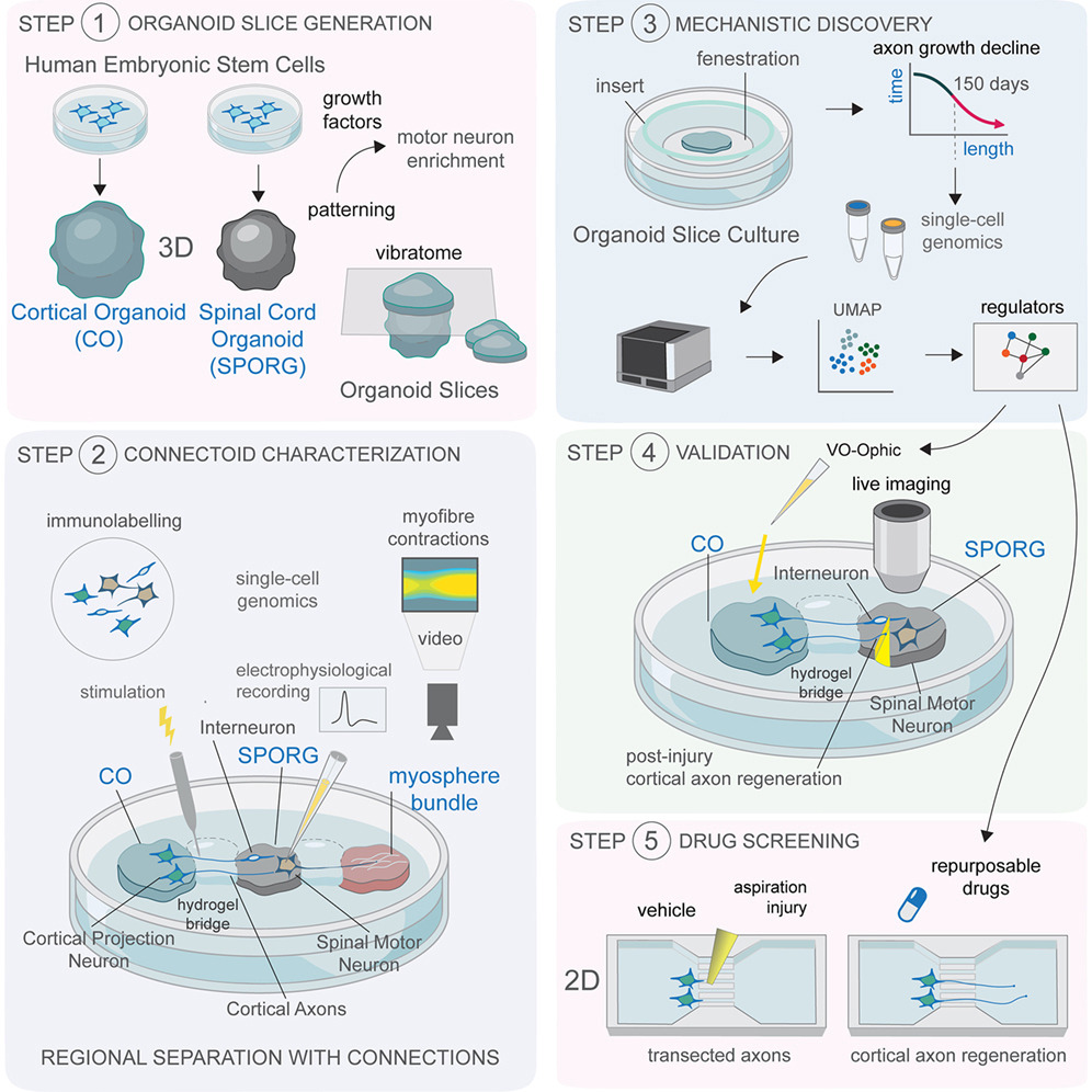
From a scientific standpoint, the most remarkable finding was that these connections formed fully functional neural circuits. These structures were capable of stimulating muscle cell contractions in the exact same manner as real movement occurs in the human body.
The researchers monitored these biological models for over a year, allowing them to identify a critical developmental window. They discovered that up to approximately day 150—the equivalent of the mid-gestational period—damaged axons retain their capacity for regrowth. Beyond this chronological threshold, the rate of regeneration drops sharply.
This finding demonstrates that the loss of regenerative capacity is not driven solely by external trauma or environmental factors; rather, it is a process hardwired into the genetic programming of neurons as they mature.
The Genetic Switch
A pivotal scientific breakthrough was the identification of the gene network that acts as a biological “switch,” suppressing axonal growth in mature neurons. When researchers artificially blocked key components of this network, the neurons regained their ability to regenerate. This milestone lays the groundwork for an entirely new therapeutic strategy aimed at reactivating growth processes within damaged nerve fibers.
In their search for substances that could influence this genetic mechanism, the scientists identified lynestrenol. This hormonal medication has long been approved in medicine for contraception and treating menstrual cycle disorders. When tested on damaged neurons, the drug significantly accelerated axonal regrowth. While this is not yet a ready-to-use clinical protocol, the precedent demonstrates that repurposing existing, approved medications could provide a major boost to nervous system repair.
Organoids: An Alternative to Animal Models
The authors note that post-injury rehabilitation faces barriers beyond genetics, such as inflammation and scar tissue development. However, directly managing neuronal mechanisms represents a fundamental step forward.
Intriguingly, relatively “young” neurons maintained their growth trajectory even within aggressive environments that typically block regeneration, underscoring the critical importance of targeting the right developmental window.
Unlike animal models, which often fail to accurately replicate human-specific biology, stem cell-derived organoids offer a high-fidelity platform for studying diseases and testing new drugs. Notably, this methodology is already actively utilized across various medical fields—ranging from liver regeneration to the study of gastrointestinal pathologies and early pregnancy phases.
Dr. Lakatos believes there is still a long research path ahead before reaching clinical practice, but the current results are highly promising. If scientists can ultimately refine methods for safe axonal growth and the proper restoration of neural connections, conditions previously deemed incurable – including severe spinal cord injuries—could become far more manageable in the future.
Source: University of Cambridge
