


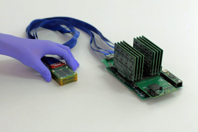
MIT scientists have developed a miniature ultrasound system that is radically changing the early diagnosis of breast cancer. The new device is so small that it can be used both in a doctor’s office and at home. This technological advantage is particularly crucial for high-risk patients.
Between 20% and 30% of breast cancer cases are “interval cancers”—tumors that develop in the period between scheduled mammograms. These tumors are often more aggressive. When diagnosed at an early stage, the patient survival rate is nearly 100%, but in later stages, this figure drops to 25%.
Current ultrasound machines are bulky, expensive, and require highly skilled technicians to operate. MIT’s new system breaks down these barriers.
How does the system work?
The new system consists of two main components, the first of which is a compact ultrasound probe. Traditional ultrasound machines use sensors arranged in a single line, capturing only flat, two-dimensional “slices” of tissue. This often requires manual movement of the device and carries the risk of missing small details.
In contrast, the device created by MIT features sensors arranged in a square sparse array. This allows the system to perceive depth from multiple angles simultaneously—without applying pressure or deforming the tissue—and directly create a full, volumetric 3D image where no spot remains hidden.
The second vital component is the data-processing motherboard, which is slightly larger than a smartphone. It costs only $300 to manufacture, consumes very little power, and provides real-time imaging via a standard laptop, making the entire system fully portable.
The system connects to a laptop and displays 3D images in real time. Unlike traditional machines, this probe does not require heavy pressure on the tissue. This mechanism reduces image distortion and allows cysts or growths to be seen in their natural state.
Future Outlook
Scientists are working on an even smaller version of the system, which will be the size of a fingernail and connect to a smartphone. There are also plans to develop a dedicated app that, with the help of Artificial Intelligence (AI) algorithms, will guide the patient on exactly where to place the probe to obtain the best image.
The technology is currently undergoing clinical trials at MIT and Massachusetts General Hospital (MGH). Ultimately, the scientists’ goal is to make ultrasound screenings as accessible as possible for everyone.
