


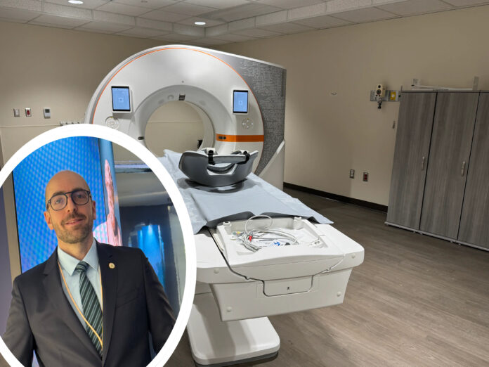
Photon-Counting Computed Tomography (PCCT) – A Revolution in Medical Imaging and Cardiovascular Diagnostics
Photon-Counting Computed Tomography (PCCT) implies a completely new form of computed tomography, a new technological breakthrough, and the rapid resolution of many such medical problems that we could not even dream of until now. If we want to understand what this technology represents, it is essential to have a good understanding of the specifics of cardiovascular diseases and how crucial good, high-quality visualization of the heart and its structures is.
Along with this topic, the reader of Medscriptum will also be introduced to the comments of Paul Maurovich-Horvat, Head of the Medical Imaging Centre in Budapest and Chairman of the Department of Radiology at Semmelweis University, who is considered one of the leading specialists of this technology worldwide.
Cardiovascular Diseases
Cardiovascular diseases (CVD) represent the leading cause of mortality and deterioration of the quality of life in the modern world, causing the death of approximately 19.8 million people annually. The primary cause of this global burden is coronary artery disease (CAD / IHD), which is characterized by atherosclerotic plaquing, blockage, and stenosis of the lumen of the heart’s main coronary arteries. The narrowing of the internal diameter of the blood vessel threatens adequate myocardial perfusion, restricts oxygen exchange at the cellular level, and manifests clinically as stable angina, acute coronary syndrome (ACS), or asymptomatic (silent) myocardial ischemia. Notably, the risk of recurrent ischemic events is 6 times higher in patients who have survived a myocardial infarction.
Against this background, radiological diagnostics, specifically coronary computed tomography angiography (CCTA), is of decisive importance for selecting the optimal treatment strategy. However, computed tomography operating on the basis of traditional Energy-Integrating Detectors (EID) frequently encounters fundamental physical limitations. Such is the case with the so-called “Blooming Artefacts” generated around calcified plaques, which leads to an overestimation of the degree of stenosis. It is precisely in response to these challenges that a massive technological leap was made in the field of radiology in the form of Photon-Counting Computed Tomography (PCCT), which radically changes the standards of medical imaging.
Physico-Technical Aspects: From Traditional CT to PCCT
The history of computed tomography dates back to 1963, with the theoretical papers of Allan Cormack, and the creation of the first clinical EMI scanner by Godfrey Hounsfield in 1971, for which they earned the Nobel Prize. Since then, the equipment has evolved continuously, but the operating principle of the detectors remained unchanged for a long time. Traditional scanners utilize a two-step process with Energy-Integrating Detectors (EID): X-ray photons first hit a scintillator, where they are transformed into visible light, and subsequently, photodiodes convert this light into an electrical signal. This approach sums up the entire incoming energy and, consequently, lacks the ability to differentiate the energy levels of individual photons.
Professor Paul Maurovich-Horvat notes: “Photon-counting computed tomography is the most advanced among the tomographic technologies existing today. The main difference compared to traditional CT scanners lies in the detector technology. The detectors are capable of counting individual photons of X-ray energy, which allows us to obtain significantly better spatial resolution while using lower doses of radiation for the patient.”
Unlike traditional systems, PCCT completely eliminates the scintillation layer. It utilizes semiconductor materials (for example, Cadmium Telluride – CdTe), where X-ray photons are directly converted into electrical impulses. Every individual photon is recorded separately, indicating its precise energy characteristic, which reduces or completely eliminates electronic noise (artifact variability) and ensures zero loss of photons. Sorting photons by energy (Energy Binning) allows for the simultaneous acquisition of multi-energy data without additional radiation, making it possible to characterize tissues at the molecular and atomic levels.
Comparative Advantages and Clinical Resolution over Conventional Tomography
Photon-counting computed tomography possesses several critically important advantages compared to traditional multislice CT scanners. First and foremost, this is reflected in ultra-high spatial resolution. Since the pixel design in PCCT detectors does not require the presence of reflective septa, which are used for pixel isolation in conventional devices, it became possible to sharply reduce the size of the detector elements. Professor Paul Maurovich-Horvat pays special attention to this achievement and emphasizes that with this technology, it is possible to achieve a reduction in image slice thickness to less than 0.2 millimeters, which is a massive step forward compared to conventional CT systems that have half-millimeter resolution.
For nearly two decades, the spatial resolution of CT scanners remained relatively stable, so this new generation of detectors simply represents the everyday reality of the future in the development of the entire field. The second important factor is the elimination of blooming artifacts. In traditional CT, high-density structures, such as small calcium plaques or metal stents, blur and appear enlarged due to light scattering. The high resolution and spectral filtration of PCCT reduce this effect, allowing the radiologist to realistically assess artery lumen stenosis and avoid false-positive diagnoses. Added to this is a dramatic reduction in the radiation dose. The optimization of ionizing radiation is a top priority for patient safety. Studies have shown that the dose efficiency of PCCT allows the radiation dose to be reduced by up to 77% in coronary calcium scoring protocols, for example.
Also noteworthy is the possibility of Virtual Non-Contrast imaging (VNC). Thanks to spectral data, specialized algorithms can digitally subtract iodine contrast from the image during the post-processing stage. This reduces the number of required scanning phases and lowers the radiation dose by an additional 19%.
Pathomechanisms of Coronary Artery Disease (CAD) and the Role of PCCT in Plaque Analysis
Understanding the pathophysiology of atherosclerosis is essential for evaluating the clinical value of PCCT. The process begins with damage to the intima of the blood vessel, the migration of monocytes into the subendothelial layer, and the accumulation of oxidized low-density lipoproteins (LDL), which forms the so-called “foam cells.” As a result of a chronic inflammatory response, cytokine release, and smooth muscle cell proliferation, an atherosclerotic plaque is formed, consisting of a necrotic lipid core and a fibrous cap. Over time, the fibrous cap becomes calcified.
From a clinical perspective, the greatest threat is posed not only by stable plaques but also by so-called vulnerable or high-risk plaques (HRP), the rupture and subsequent thrombosis of which become the immediate cause of acute infarction.
Traditional coronary CT angiography (CCTA) often fails to accurately differentiate the internal structure of the plaque. With the help of PCCT, however, it is possible to clearly distinguish the lipid core, fibrous tissue, and microcalcifications based on their unique spectral attenuation (Hounsfield Units – HU). This allows coronary risk to be adequately assessed and helps prevent inappropriate or, conversely, delayed invasive treatment. This is particularly crucial, for example, in patients with type 2 diabetes mellitus, where due to autonomic neuropathy, ischemia often progresses asymptomatically and manifests directly as a fatal cardiac event.
Professor Paul Maurovich-Horvat confirms that in clinical practice, these capabilities yield revolutionary results. In his words, since photon-counting CT automatically provides spectral imaging, specialists can obtain unprecedentedly accurate visualization of coronary plaques. At the same time, the possibility emerges to detect previously hidden pathologies, such as microhemorrhages or very small fractures, the identification of which would otherwise be practically impossible and extremely difficult.
Technology Accessibility, Pricing, and Clinical Experience
Despite this technology being immensely powerful, its widespread implementation in real medical practice is still associated with certain barriers. As Professor Paul Maurovich-Horvat points out, the system is currently quite expensive and available only in specialized, large medical facilities. In Budapest, at their leading institution, three photon-counting CT scanners are currently operational, serving as the primary imaging systems. Similarly, when talking about the same technology at the Department of Radiology in Innsbruck, Austria, the leading Professor Gudrun Feuchtner told us the same thing: that it is a highly expensive luxury and they can be seen in only a few centers worldwide, including Innsbruck.
Although they are used for many types of examinations, including particularly effectively for cardiological imaging and emergency diagnostics, existing resources currently allow only for scientific research and the discussion of particularly complex cases. The professor expresses strong hope that the situation will change in the near future. According to his forecast, as more manufacturers create their own photon-counting systems, competition will drive the price down. He is confident that within five years, this technology will become much more accessible to a wide network of clinics, allowing radiology to abandon the old, energy-integrating standards.
I also have the privilege of a very close personal interaction with this technology, which gives me the opportunity to compare images provided by conventional CT angiography and photon-counting technology myself. The difference is truly astonishing. PCCT represents an undeniably unrivaled step forward not only in radiology but also in clinical medicine in general. By overcoming the physical barriers of traditional CT—even by reducing the slice thickness down to 0.2 millimeters—we are given the opportunity to obtain images with a much higher frame rate.
By minimizing artifacts, sharply reducing the patient’s radiation load, and enabling the use of a much smaller volume of contrast material, we are given the opportunity to further relieve patients of the risk of mortality and diminished quality of life. This technology lays the foundation for personalized cardiology. Early diagnosis of coronary artery disease, timely identification of high-risk vulnerable plaques, and precise planning of interventional treatment using PCCT will significantly reduce global mortality and improve the quality of life for millions of people.
