


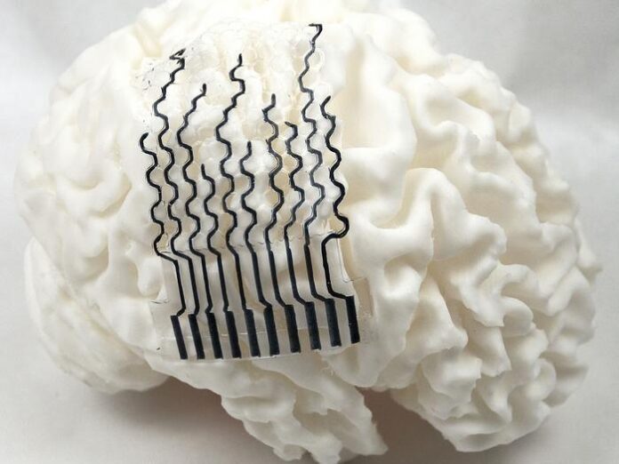
Scientists at the University of Pennsylvania have developed innovative, soft bioelectrodes tailored directly to the unique structure of a patient’s brain. According to the researchers, unlike traditional rigid and standard-shaped sensors, 3D-printed electrodes conform perfectly to the brain’s surface, opening entirely new possibilities for monitoring and treating neurodegenerative diseases.
According to the study published in the journal Advanced Materials, the surface of the human brain consists of folds and grooves (gyri and sulci) that are unique to each individual. Existing neurointerfaces fail to account for these differences; as a result, standard electrodes cannot establish full contact with the brain and can sometimes even damage soft tissues.
To solve this problem, scientists created detailed 3D brain models based on MRI scans of 21 patients and manufactured individualized bioelectrodes for each. The electrodes are primarily made of water-rich hydrogel, which gives them their flexibility.
Of particular interest is the cellular, honeycomb structure of the electrodes. Thanks to this design, the device is much softer and lighter while maintaining mechanical strength and high sensitivity for signal transmission. Additionally, the use of 3D printing minimizes production time and costs.
Laboratory tests have confirmed that these innovative electrodes establish a nearly perfect connection with brain signals. Importantly, despite the close contact, they do not interfere with the circulation of fluid around the brain—one of the main drawbacks of traditional sensors.
A 28-day observation period in animal models showed no immune response to the hydrogel, nor did the sensitivity of the electrodes decrease. In the future, the project authors plan to use this technology not only for monitoring but also for the clinical treatment of specific neurological diseases.
