


Wound infections have become one of the most alarming and growing threats to modern global healthcare. This critical situation is further exacerbated by the phenomenon of antibiotic resistance, which transforms even seemingly harmless injuries into life-threatening conditions. The severity of this scientific challenge is reflected in statistical data, according to which bacterial resistance to medication is already the cause of more than a million fatal outcomes annually. If current trends remain unchanged, the mortality rate could reach 10 million per year by 2050.
Complex wound infections account for a major share of this global crisis. Currently, almost half of them exhibit resistance even to powerful drugs like third-generation cephalosporins. Although beta-lactam antibiotics are still considered the “gold standard” for treating skin and soft tissue infections, aggressive pathogens (Pseudomonas aeruginosa and MRSA) utilize a powerful defense mechanism—beta-lactamases. These enzymes purposefully break down the nuclear chemical ring of the medications, thereby depriving the antibiotics of their ability to fight bacteria. This process fully neutralizes the efficacy of the drug and calls the clinical outcome of the treatment into question.
The situation is further aggravated by the indiscriminate use of passive dressings, which provide a continuous stream of low-concentration antibiotics to the healthy microflora of the skin. When the concentration of an antibiotic is insufficient to fully destroy the bacteria, the microorganisms learn new survival strategies. Consequently, instead of providing treatment, we facilitate an unintended process of “training” the bacteria, making them completely impervious to medications.
Researchers at Brown University have responded to this complex challenge facing the healthcare system with a high-tech “smart hydrogel.” This material fundamentally changes standard wound care methods and establishes a targeted, “on-demand” defense mechanism against pathogens.
How was the Smart Hydrogel Created?
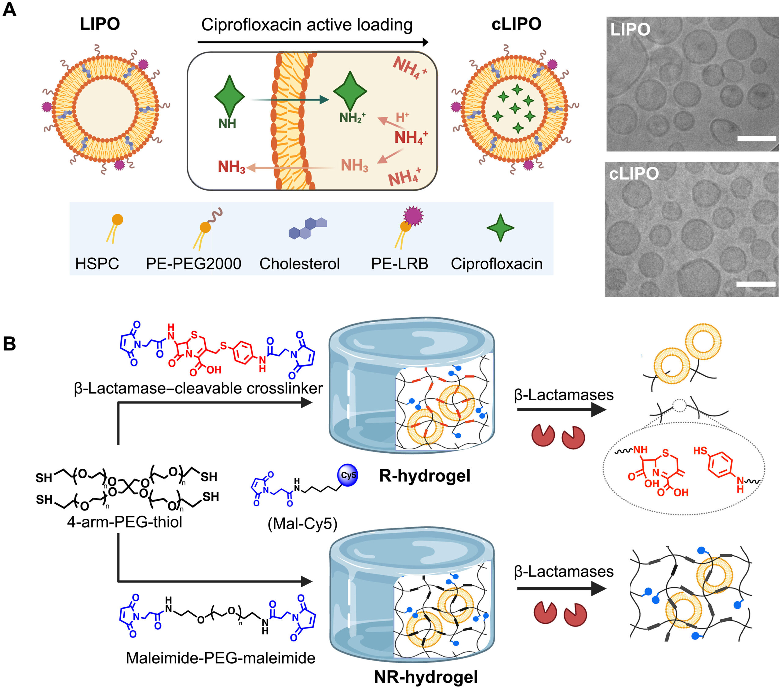
The researchers created the innovative hydrogel from long, hydrophilic (water-loving) polymer chains that are linked together by special cephalosporin “bridges.” These connecting segments mimic the structure of beta-lactam drugs.
The operating principle of the system is calculated directly based on the bacterial defense mechanism. As soon as the beta-lactamases secreted by bacteria attack these connecting bridges, the entire structure of the hydrogel begins to break down. As for the actual therapeutic substance, ciprofloxacin (an antibiotic from the fluoroquinolone group that inhibits bacterial DNA replication) is initially placed inside liposomes. These nanospheres (~105 nm) successfully prevent the premature activation and non-targeted release of the drug. The homogeneous distribution of liposomes throughout the volume of the gel ensures a precise and even release of the therapeutic dose.
It is also noteworthy that these innovative materials react instantaneously to specific biological signals (changes in pH, reactive oxygen species (ROS), or bacterial enzymes). For example, a pH-sensitive design utilizes acid-labile bonds that break down on their own in the acidic environment characteristic of infected tissues. Similarly, ROS-triggers are activated directly at the foci of inflammation, while enzyme-responsive gels identify specific proteins of pathogens.
In early attempts, scientists used lipases or proteases, but these overlapped with natural human enzymes, creating a risk of premature and non-targeted drug release. Since beta-lactamases are not produced by the human body, the medicine is activated only during a bacterial attack. It is this exclusive sensitivity to bacterial enzymes that makes the system ready for error-free and targeted action.
Structural Composition of the Hydrogel
To create the new material, scientists combined special polymer chains (4-arm PEG-thiol polymers) and cephalosporin molecules (derived from 7-amino-3-chloromethyl-3-cephem-4-carboxylic acid). Despite this complex chemical composition, the resulting gel is not inferior in strength or structure to conventional medical dressings. Upon observation of the process (using fluorescent markers—Cy5 and Rhodamine B), it was established that 88% of the therapeutic substance is reliably housed directly within the gel.
The exceptional stability of the system played a decisive role in the success of this technology. Under laboratory conditions (at pH 7 or 4), no spontaneous leakage of the antibiotic was recorded over seven days. This double barrier—the gel degradable by enzymes and the liposomes existing within it—reliably ensures the release of the preparation only when there is an actual need, so-called “on-demand.”
Laboratory Phase of the Research
The functional capabilities of the hydrogel were successfully tested within the scope of an in vitro study. During the experiment, responsive gels (R-hydrogels) fully dissolved in just 6 hours when acted upon by specific enzymes from P. aeruginosa. Under the same conditions, non-responsive control samples (NR-hydrogels) fully maintained their initial stability. Fluorescent analysis detected gel fragments and released liposomes in the environment. The study showed that the concentration of ciprofloxacin reached 1.5 μg/mL in 30 minutes, which is five times higher than the minimum inhibitory concentration (MIC) required to destroy the pathogen.
The outstanding selectivity of this technology became even more apparent during the enzymatic testing phase. Various enzymes common in wound environments (collagenase, hyaluronidase, lipase, elastase, and phospholipase) were unable to break down the gel structure over 72 hours. Hydrogen peroxide, as an imitator of inflammatory processes, also had no effect on the system. However, penicillinases from Bacillus cereus caused the instantaneous breakdown of the gel, clearly confirming its sensitivity to various groups of beta-lactamases.
Microbiological analysis confirmed that the degradation of the hydrogel is directly dependent on the enzymes secreted by bacteria. On agar plates where colonies of P. aeruginosa Xen41 were present, the dressing was fully absorbed, while it exhibited complete stability toward strains lacking the enzyme (S. aureus Xen29). This fact confirms once again that without the proper biological signal, the system reliably maintains stability and strength.
The experiment demonstrated that the substances produced as a result of the material’s breakdown do not pose a threat to living cells. During the testing process, the activity of mouse fibroblasts remained unchanged, and human red blood cells fully maintained their structural integrity. Such biosafety is a prerequisite for the future practical implementation of the technology.
Results from the Mouse Model
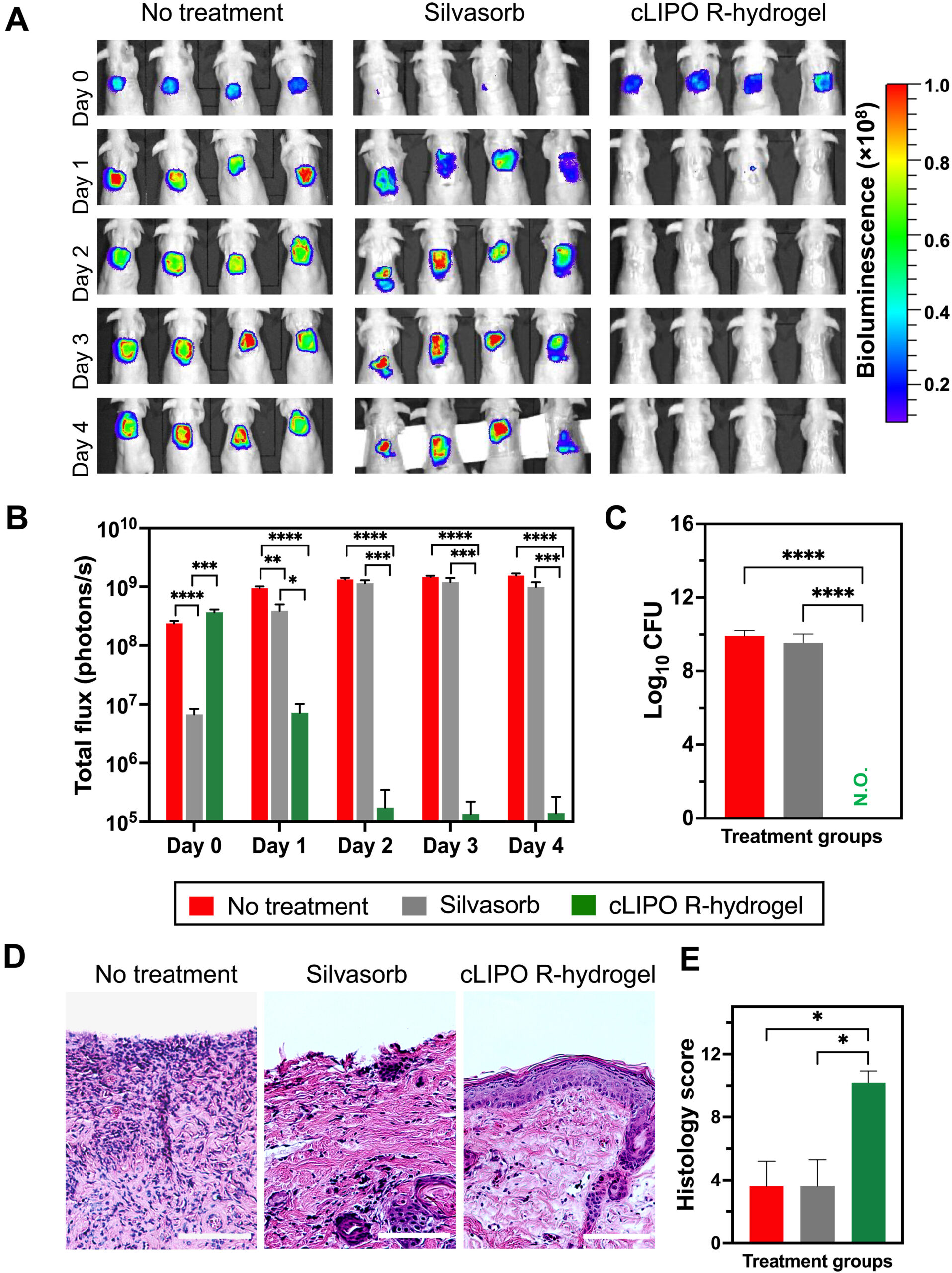
To test the efficacy of the technology, the researchers used a mouse skin injury model that mimicked a real infected wound. During the experiment, a high concentration of P. aeruginosa was introduced into the wound, after which the spread of bacteria was observed using the bioluminescence method for 4 days. Initial observations conducted on drug-free gels showed that only the responsive (R) gels degraded, which led to the release of the liposomes within them.
For comparison, Silvasorb (a modern silver-based clinical standard) sharply reduced the number of bacteria in the first 2 hours but allowed them to grow again the following day. In contrast, the antibiotic-loaded smart hydrogels brought the number of pathogens to a minimum by the second day. By the fourth day, the bacterial count in tissues treated with the R-gel equaled zero, while infection remained at a high level in the other groups.
From a clinical perspective, the quality of healing proved to be optimal. Histological analysis revealed full restoration of the skin cover and minimal damage in mice treated with the R-gel. Furthermore, immunofluorescence confirmed a sharp reduction of inflammatory processes at the site of the injury.
Porcine Skin Burn Model and Prevention of Resistance
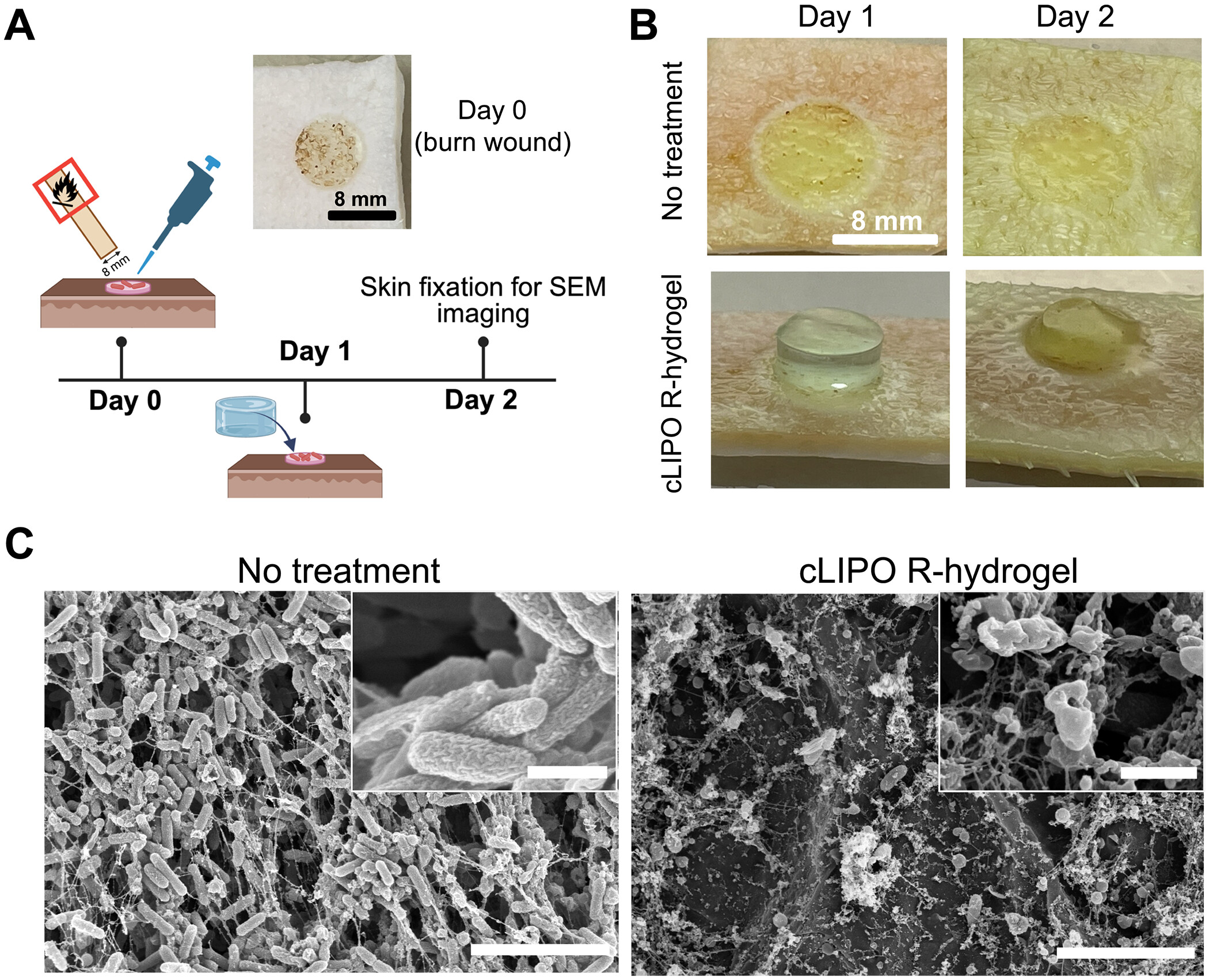
Research conducted on a porcine skin burn model tested the practical effectiveness of the technology once again. Full breakdown of the “smart” (R) gels in infected wounds was recorded in just one night. Electron microscopy showed that instead of solid biofilms, only bacterial remnants remained in the injured area.
The resistance analysis proved to be particularly significant: a simulation of 10-day prophylactic use showed that the sensitivity of pathogens to the drug did not change at all. For comparison, the use of free ciprofloxacin in small doses caused an eightfold increase in resistance by the fourth stage.
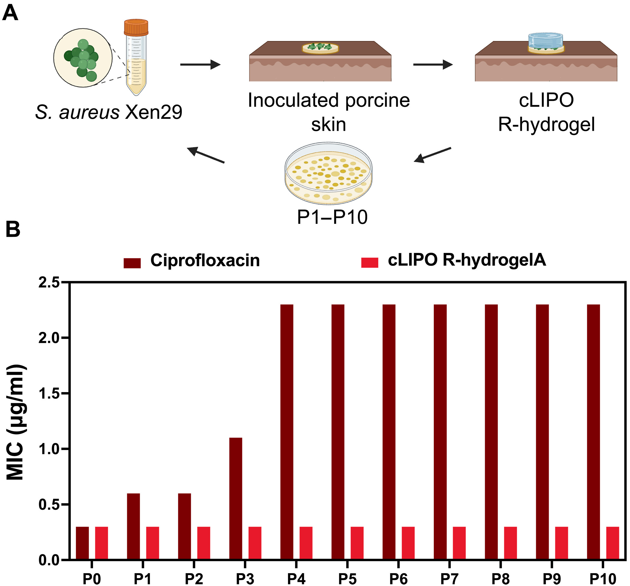
Thus, the strict control of the hydrogel successfully prevented the premature release of the drug and selective pressure on the bacteria. These results confirm that the new technology effectively cures infection and, at the same time, hinders the development of antibiotic resistance.
Advantages, Limitations, and the Future Path
This hydrogel significantly outperforms passive means, such as silver-containing dressings, in its operating principle. Unlike traditional methods that release a drug constantly and uncontrollably, “smart” (R) hydrogels destroy an infection only at the necessary moment with a targeted strike.
The enzymatic levels used in the study fully correspond to real clinical indicators. Since the human body does not produce beta-lactamases, the technology ensures high specificity and safety. The absence of toxicity and the prevention of resistance further strengthen confidence in this innovation.
Despite the success achieved, certain scientific challenges remain. In current studies, scientists used individual strains, whereas real wounds are often characterized by a diverse, polymicrobial nature. The next stage of work requires mixed models, genetic sequencing, and combination antibiotics.
The future development of the project involves fighting chronic infections and testing on large animals. To confirm the platform’s efficacy, further research with bacterial isolates obtained from real patients is planned. Along with this, refinement of scalability, self-cost, and storage stability is necessary. However, for this stage, this approach offers hope for the implementation of a completely new standard for wound treatment.
Source: Science
