


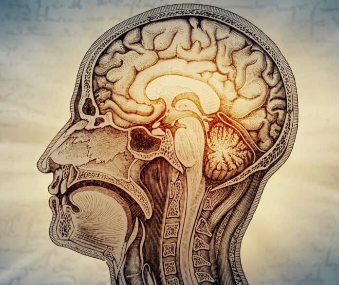
According to the long-held view, all regions of the brain developed from a single common origin during the early stages of embryonic development. However, researchers at Stanford University have discovered that two distinct progenitor cell populations participate in brain development. Although they emerge simultaneously, their genetic programs differ, driving the development of distinct regions of the brain.
According to the study, brain development merges two ancient nervous systems: one linked to vital functions, including breathing and heart rate, and the other driving cognitive abilities, abstract thinking, and self-awareness.
But how do these two distinct trajectories emerge during embryonic development?
The neural ectoderm is an early embryonic tissue from which the brain and nervous system arise. Experiments on mouse embryos—in which scientists traced cellular developmental trajectories—demonstrated that early in development, still during the gastrulation phase, two progenitor populations of neural ectoderm emerge concurrently: the anterior and posterior neural ectoderm.
The anterior neural ectoderm gives rise to the forebrain and midbrain, while the posterior neural ectoderm drives the development of the hindbrain. Different genes are activated in each: Otx2 in the anterior neural ectoderm and Gbx2 in the posterior. This divergent genetic activity steers their development along separate trajectories.
The divergence is not limited to gene activity alone. The researchers also examined chromatin—the structure inside the cell nucleus in which DNA is packaged. Chromatin organization dictates which genes a cell can activate or repress. It turned out that the anterior and posterior neural ectoderm exhibit distinct chromatin architecture corresponding to their predetermined developmental fates from this early stage. Thus, the future destiny of these cells is determined even before specific brain regions physically form.
How did this discovery help researchers generate neurons?
One of the study’s key milestones was deriving hindbrain motor neurons from human pluripotent stem cells—specifically, cranial motor neurons characteristic of hindbrain rhombomeres 5 and 6, which are involved in facial mimicry and swallowing. Previously, generating this specific subtype of neurons in the laboratory posed a significant challenge. However, deciphering this early developmental genetic program enabled researchers to generate cells that accurately recapitulate the electrical impulses and functional hallmarks of these motor neurons.
Furthermore, the finding opens new avenues for studying neurodegenerative disorders such as spinal muscular atrophy (SMA) and amyotrophic lateral sclerosis (ALS). Since motor neurons are selectively damaged in such pathologies, laboratory analogs provide a platform to observe disease mechanisms and screen potential therapeutic compounds.
Remarkably, scientists identified a similar pattern across evolutionarily distant organisms—including fish, birds, and hemichordates (acorn worms), marine invertebrates that share a deep common ancestor with humans. They hypothesize that these two progenitor populations have been evolutionarily conserved for approximately 550 million years.
