


In recent years, neurosurgery has undergone rapid development, and one of the most innovative directions has become minimally invasive approaches that allow brain surgery to be performed without traditional craniotomy. A particularly important breakthrough in this field was recently reported in the United Kingdom, where a brain aneurysm was treated via the eye socket for the first time using a transorbital approach.
The operation was performed at Leeds Teaching Hospitals NHS Trust, one of the leading medical and university hospital networks in the country. This center is known for its high-level neurosurgical expertise and its involvement in the development of advanced surgical techniques. In this case, surgeons used a completely new anatomical “corridor” — the orbit of the eye — as an access route to deep intracranial structures.
The patient was a 61-year-old man diagnosed with a cerebral aneurysm. Aneurysms are pathological dilatations of blood vessel walls that can rupture and lead to life-threatening subarachnoid hemorrhage. Traditionally, such cases are treated either by open neurosurgery (craniotomy) or endovascular techniques such as coil embolization. However, in this case, the surgical team opted for a more innovative approach — a transorbital endoscopic technique.
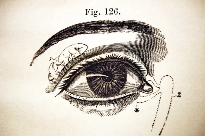
The main principle of this method is the use of a natural anatomical space. The orbital cavity serves as a relatively direct biological corridor toward the skull base. During the procedure, a small, carefully planned incision was made in the eyelid region, after which an endoscopic system was inserted through the orbit. This system includes a high-resolution camera, a light source, and microsurgical instruments, allowing real-time visualization of the surgical field on a monitor.
One of the most challenging aspects of the procedure was the extreme anatomical sensitivity of the orbit. It contains critical structures such as the optic nerve, extraocular muscles, arteries, and veins. Any minor injury can result in vision impairment or neurological complications. Therefore, preoperative planning included high-resolution MRI and CT imaging as well as 3D navigation systems to precisely map the surgical pathway.
The main advantage of the transorbital approach is the significant reduction in surgical trauma. Unlike craniotomy, it avoids large bone removal, thereby reducing infection risk, postoperative pain, and recovery time. Patients may benefit from faster rehabilitation and shorter hospital stays.
However, this technique is still considered highly specialized and is used only in selected cases where the aneurysm location allows safe access through the orbit. According to experts, this approach represents a potential paradigm shift in neurosurgery and may, in the future, become a standard alternative for certain skull base vascular pathologies.
