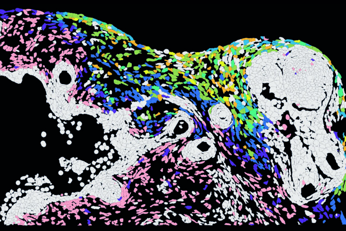
Herpes simplex virus 1 is best known for causing cold sores, but researchers are still working to understand how it moves through human skin and why infection can occasionally become serious.
A new study published in Science Advances used human hair-bearing skin organoids to trace HSV-1 infection cell by cell. The model, developed by researchers at the Max Delbrück Center and University Medical Center Hamburg-Eppendorf, contains epidermal cells, dermal cells, hair follicles and nerve-like cells, making it more similar to real skin than a flat cell culture.
A 3D view of infection
The researchers combined imaging with spatial transcriptomics, a technique that measures gene activity in individual cells while preserving their location within tissue. This allowed them to follow the virus as it spread through different layers of the organoid.
HSV-1 did not infect every skin cell type in the same way. Dermal fibroblasts carried large amounts of virus early in infection. Basal keratinocytes in the lower epidermis and several hair-follicle cell types carried relatively less virus, even when infected cells were nearby. The virus reached hair follicles and sensory neurons later, suggesting that these cells may have some intrinsic resistance.
The findings do not mean that HSV-1 always follows this exact route in people. They show how infection progressed in this particular three-dimensional skin model, providing a more realistic setting for studying viral spread than conventional two-dimensional cultures.
Cells send signals to their neighbours
The study also examined how cells at the leading edge of infection responded. At the infection front, dermal fibroblasts activated IER3, a gene known to respond to HSV-1. Basal keratinocytes increased expression of IRF3, a regulator of antiviral defences.
The researchers found that IRF3 was increased not only in infected keratinocytes but also in neighbouring uninfected cells. This pattern is consistent with paracrine signalling, in which infected cells influence nearby cells and help prepare them for an antiviral response.
The team also identified a spatially coordinated inflammatory response involving tumour necrosis factor family signalling. TNFSF9, also known as 4-1BBL, was induced in papillary fibroblasts, while TNF-alpha was induced in keratinocytes. Laboratory tests confirmed that these cytokines and the TNF-alpha-responsive chemokines CXCL2 and CXCL10 were secreted.
Interferon-beta reduced viral replication in the model, whereas TNF-alpha and 4-1BBL did not. This suggests that their main role may be to organize immune-cell recruitment and activation rather than directly suppress the virus.
Why the model matters
HSV-1 usually infects sensory nerve cells in the face, where it can remain dormant. In rare cases, it can travel to the central nervous system and cause life-threatening brain inflammation. Researchers are also investigating whether its lifelong persistence in the nervous system may contribute to nerve-cell damage, including possible links to Alzheimer’s disease.
The organoid model does not yet include immune cells or blood vessels, so it cannot fully reproduce the human immune response to infection. Future models that add these components could help researchers study how the body controls HSV-1 before it reaches nerves, why some cells slow viral replication while others support it, and how infection-related inflammation damages tissue.
For now, the study provides a detailed map of how HSV-1 can move through a complex human skin-like tissue—and shows that the response to infection is not uniform, but highly dependent on cell type and location.
Source: Max Delbrück Center