



Worldwide, cardiac arrhythmia affects every third adult. It sharply increases the risk of stroke, heart failure, and sudden death. The severity of the condition is also highlighted by the economic factor; disorders such as atrial fibrillation (AF) and ventricular tachycardia (VT) cost the European healthcare sector alone more than 13.5 billion euros annually.
Despite these scales, existing therapeutic approaches still face significant challenges. On one hand, drug treatment is often ineffective and characterized by severe side effects. On the other hand, catheter ablation, a relatively radical treatment method, is often powerless in cases where arrhythmic foci are hidden in the deep, intramural layers of the heart wall. It is precisely this diagnostic “invisibility” that causes the exact localization of the problem to often remain unachieved despite invasive interventions.
In response to this challenge, researchers from the Polytechnic University of Valencia (UPV) and Corify Care have created a revolutionary method: Global Volumetric Mapping. This innovation allows for a complete, three-dimensional image of all four chambers to be obtained with just a single beat of the heart.
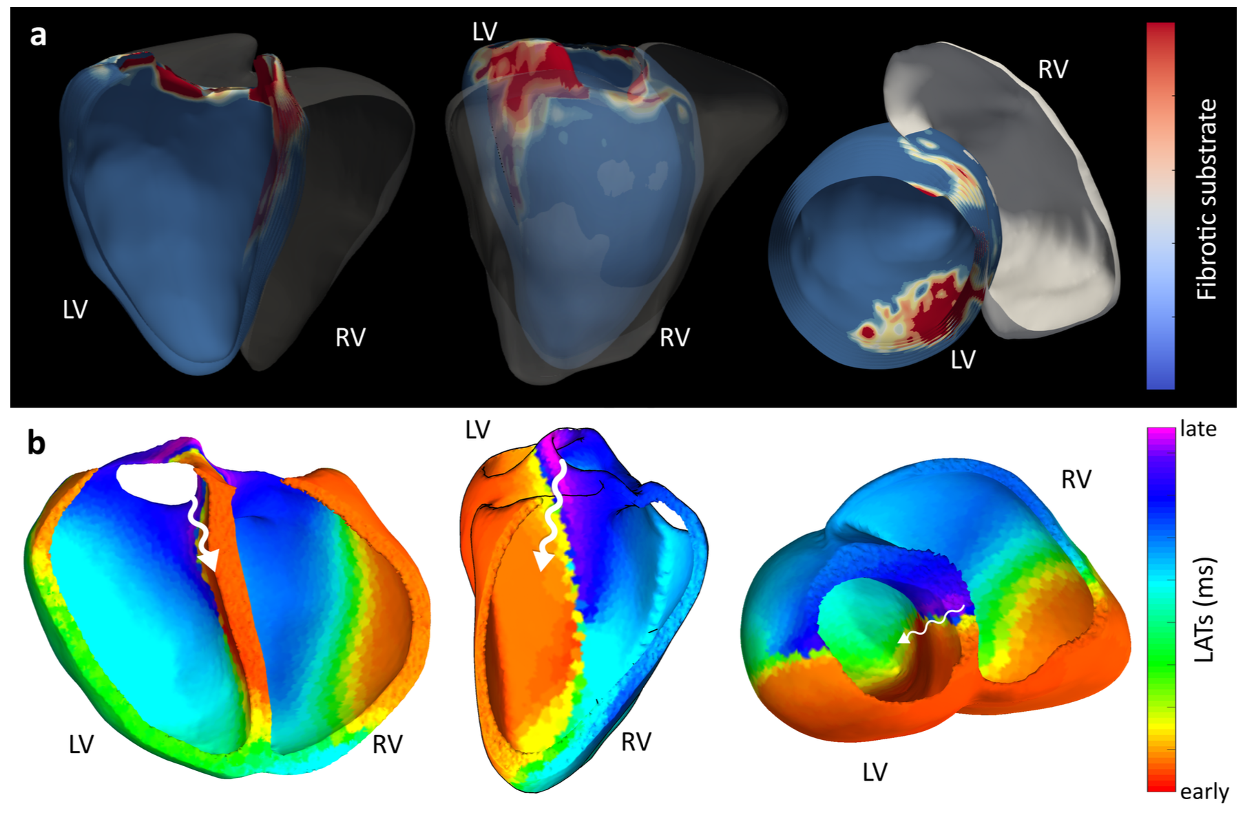
The main advantage of the technology is that it shows, in real-time, the pathways of arrhythmia propagation that pass through the heart walls and septa. These are precisely the details that were practically impossible to see with previously existing tools.
The Working Principle of ACORYS
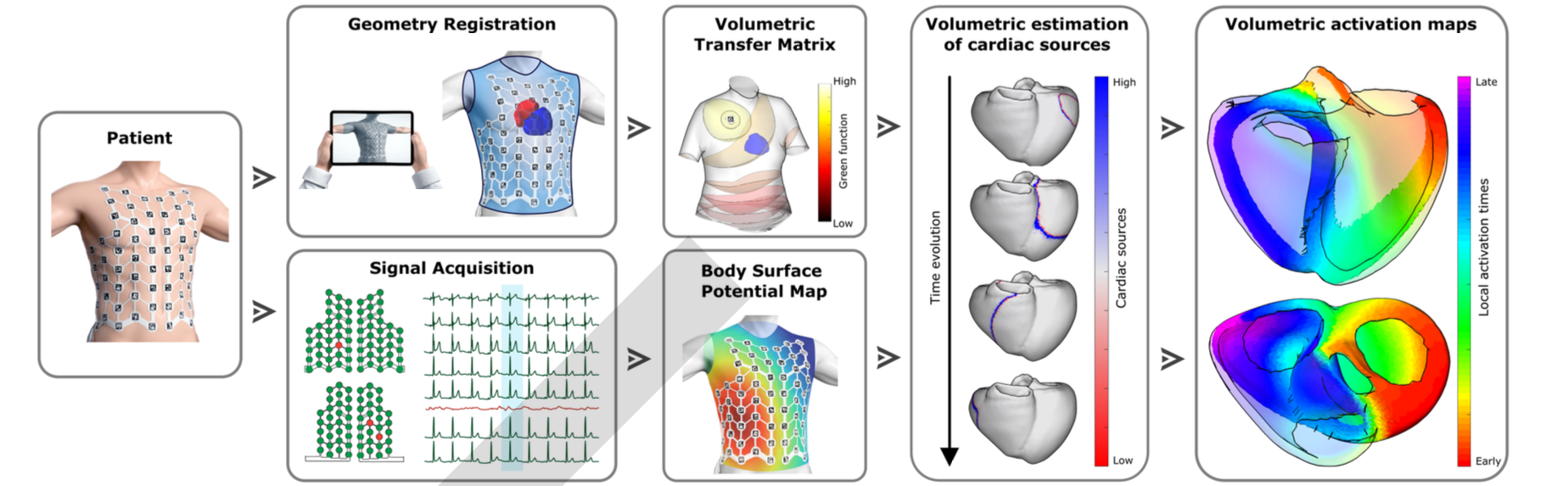
The working principle of the ACORYS system is simple and effective: the patient wears a special, 128-electrode vest that captures heart signals from the body surface. With the help of photogrammetry and mathematical modeling, the system instantaneously creates an individual 3D model of the body and the heart.
From Simulation to Clinical Practice
Computer modeling demonstrated the technology’s high accuracy. In cases of heart septum pathology, traditional methods can produce errors of up to 30 millimeters, often leading to misdiagnosis, whereas the new volumetric approach reduced this error by half.
Clinical trials in patients aged 50 to 80 confirmed these results. The system precisely identified foci of ventricular premature beats and clearly visualized the accessory pathways characteristic of Wolff-Parkinson-White (WPW) syndrome.
The technology proved especially valuable for managing conduction disorders such as Left Bundle Branch Block (LBBB), where uncoordinated chamber contractions reduce cardiac efficiency. Its 3D visualization enabled accurate planning of Cardiac Resynchronization Therapy (CRT). The system also reliably detected post-infarction scar tissue, supporting both precise diagnosis and personalized treatment.
To gain deeper insights into this breakthrough, Medscriptum reached out to Andreu Climent, PhD, CEO of Corify Care and researcher at the Polytechnic University of Valencia (UPV).
What were the primary research challenges in cardiac electrophysiology that inspired the development of the ACORYS system’s global volumetric mapping technology?
For decades, treating a heart arrhythmia has been like trying to understand what is happening in a dark football stadium using only a small flashlight. Doctors insert a catheter (the flashlight) and touch the heart wall point by point. They see the detail of where they touch, but they lose the big picture.
Our inspiration was simply to “turn on the lights in the stadium.” We wanted to end “blind” treatments. The challenge was to stop looking at fragments and start seeing the heart globally, allowing the doctor to enter the operating room with a clear roadmap. We call this Clinical Clarity.
Can you walk us through how wearing a vest for minutes gives doctors a full heart map?
Think of it as creating a personalized “Google Maps” of the heart in real-time. The process is entirely non-invasive: the patient simply wears a vest with sensors, very similar to a standard ECG but more comprehensive.
This vest “listens” to the electrical whispers of the heart from the surface of the skin. Our technology takes those whispers and, in less than 10 minutes, translates them into a high-definition 3D image. What makes us unique is that we don’t just see the “facade” of the heart; we can see through the walls, identifying the origin of the arrhythmia in a single heartbeat. It is the transition from a static photo to a “Satellite View” of all electrical activity.
What are current limits, e.g. BMI effects, VT speed, or geometry accuracy without CT/MRI, and fixes in the pipeline?
We are honest with the science: like any imaging technology, in different situations, the signal can be weaker. However, our system is smart and alerts the doctor about signal quality to always ensure precision.
The fascinating part is that what used to be a problem is now our strength. For example, in very fast and unstable tachycardias (VT) where traditional catheters fail because they don’t have time to “draw” the map, we win. Since we only need a single beat to see the full map, we can catch arrhythmias that were previously impossible to map. We are also working so that, in the near future, we won’t even need a prior CT scan, using AI to automatically recognize the unique shape of each patient’s heart.
After clinical validation in 50-80 year-olds, what’s the timeline for larger trials, FDA approval, and CRT/pacemaker integrations?
We are in what we call our “Prime Time.” After validating our technology in over 2,500 patients and publishing our results in the journal Nature Communications Medicine, we are playing in the “Champions League” of cardiology.
We are currently in the active review process with the FDA to enter the U.S. market within 2026. Furthermore, we are already working to make pacemakers much smarter (CRT therapy), allowing the doctor to see instantly how the heart responds to the device so they can adjust it for a perfect heartbeat.
Where does this lead – AI predictions, personalized pacing, or revolutionizing EP like CT did radiology? Advice for adopters?
Just as CT or MRI revolutionized medicine by allowing us to see the body’s physical structure, Corify is born to be the “Electrical Imaging” of the heart.
We are facing a true paradigm shift: for the first time, we can map the entire heart in 4D (space and time) over extended periods. The volume of data generated is so massive that it is humanly impossible to analyze every single heartbeat and signal in detail manually. This is where Artificial Intelligence plays a vital role. AI is not here to replace the doctor; it is here to act as an advanced assistant, helping clinicians “see” what was previously invisible.
AI reaches where the clinical team’s time cannot, filtering through massive datasets to deliver only the most critical, actionable insights. The ultimate goal is empowerment: ensuring that clinicians, armed with the best possible information, can make the most appropriate decisions for each unique patient.
My advice for future adopters: Don’t just see this as a new tool, but as a new standard of clarity. Just as a surgeon today wouldn’t dream of operating without a prior CT scan, it will soon be unthinkable to treat an arrhythmia without first “seeing” the heart’s electricity. It is time to stop working on assumptions and start working on evidence.
Source: nature
