


Anomalies formed during fetal development often determine the entire future life and physical capabilities of a child. Therefore, the priority of modern medicine has become to confront pathologies not after birth, but while in the womb, at the stage of fetal formation. The main essence of this approach lies in making it possible, through timely intervention, to stop the pathology before it causes irreversible damage to the organism.
Project CuRe of the University of California (UC Davis) is guided by this principle, which implies the integration of traditional fetal surgery and regenerative therapy. Data published in the journal The Lancet confirm the safety of the given combined method. While the next stage of the study aims to determine how effectively this approach helps children in moving independently and maintaining physical functions.
Spina Bifida
Spina bifida belongs to the group of congenital defects of the neural tube. The neural tube represents an embryonic structure from which the brain, spinal cord, and their surrounding tissues are subsequently formed. Under conditions of normal development, this structure is formed at an early stage and closes on the 28th day from conception. However, during the given pathology, the perfect formation or timely closure of a certain part of the neural tube does not occur, which determines defects of the spinal cord and the vertebrae of the spine.
The amniotic fluid existing in the womb chemically irritates the open nervous tissue, while constant mechanical friction damages it even further. Precisely this aggressive impact becomes the cause of those severe complications that manifest as paralysis, the accumulation of fluid in the brain, or the disturbance of the functions of internal organs.
Over time, the treatment strategy was significantly refined. If previously the surgical closure of the defect occurred only after birth for the purpose of preventing infections, a large-scale clinical study (MOMS) published in 2011 confirmed the advantage of intrauterine surgery. Fetal surgery indeed reduced the necessity of shunting against the accumulation of fluid in the brain and also improved motor skills, although the main challenge still remained unresolved.
Specifically, more than half of the operated children are still unable to move independently at the age of 2.5 years. This is explained by the fact that standard surgery only protects the spinal cord from further damage, although it does not possess the resource for the restoration of already existing nervous defects.
Why Stem Cells?
In order to also add regenerative properties to the surgical intervention, scientists stopped their choice on placental mesenchymal stem cells (PMSC). These cells ideally answered the set goals: obtaining them from donor placentas is easy, while due to their fetal origin, the risk of tumor development is also minimal.
Besides, these cells multiply quickly in the laboratory and secrete far more neurotrophic factors (BDNF, HGF) compared to analogues from the bone marrow of an adult person. Precisely these biologically active substances play a decisive role in the survival of neurons, because they effectively block inflammatory processes and protect the cells from dying.
This regenerative potential of the cells was initially confirmed by laboratory tests, which gave scientists the basis to re-verify their effectiveness on animal models. In conditions similar to human prenatal development, during fetal surgery conducted on lambs, the stem cells ensured protection from the damage of the spinal cord.
As a result of the experiment, the animals fully maintained motor functions, as well as the physiological work of the intestines and the urinary bladder. This successful model also determined the optimal dosing of the treatment: 300,000 cells are placed on every square centimeter, which are distributed on a special bio-covering (the so-called “patch”) approved by the FDA.
After the positive results obtained on in-vitro and animal models, UC Davis researchers Diana Farmer and Aijun Wang, after a 10-year search, arrived at the optimal and safest method. They refused the use of the patient’s own cells due to risks connected with biopsy and stopped the choice on donor (allogeneic) material. The final confirmation of the safety of this method laid the foundation for a large-scale clinical study— “CuRe,” which began in 2021.
The CuRe Study: From Theory to Practice
The CuRe phase one clinical study, the aim of which was the confirmation of the safety of the innovative method, began with a strict scientific protocol for the selection of participants. On the basis of criteria analogous to the “MOMS” study, pregnant women were selected according to the gestation period (19-26 weeks), the normal karyotype of the fetus, and a clear clinical picture of myelomeningocele. At the same time, to maintain the objectivity of the results, those cases were excluded from the process that contained the risk of premature birth or other concomitant pathologies.
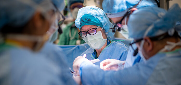
The laboratory preparation started 72 hours before the surgery represented the decisive moment of this difficult intervention. In a specialized environment, scientists checked the sterility and viability of the cells in detail, after which the biological material was placed on a bio-matrix for 24-hour incubation. As soon as it passed the final inspection, the ready cellular “patch” was transferred to the operating room, where a prepared open section of the fetal spinal cord was already waiting for the surgeons. After the appearance of the uterus of the mother under general anesthesia, the layer of pre-cultivated stem cells was placed directly on the damaged area.
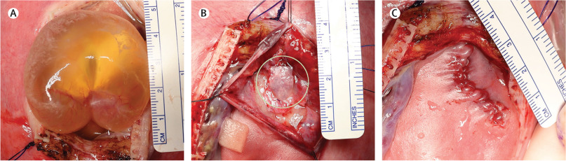
After the hermetic reconstruction of the spinal cord membrane and the skin, the operation was completed and the stage of many-week ultrasound observation on the condition of the fetus began. Within the framework of the study, the children were born on average at the 34th week of gestation by cesarean section. Despite the fact that two newborns required short-term respiratory support in the postnatal period, all of them were discharged from the hospital without dependence on oxygen, with stable respiratory function.
Study Results
The primary results of the study, which are based on six patients enrolled in 2021-2022, are hopeful in terms of the safety of the method. Postnatal magnetic resonance imaging (MRI) showed that the herniation of the back part of the brain was fully corrected in all newborns, which constitutes one of the primary clinical goals of fetal surgery. At the same time, a detailed medical examination confirmed that in no case was infection, wound opening, liquorrhea, or any kind of complication associated with stem cells recorded, including neoplastic processes.
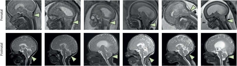
By visual inspection, the reconstructed areas did not differ from cases treated with traditional surgery, which indicates that the addition of cells did not increase the procedural complexity. It is also important that until the discharge from the hospital, not a single child required shunting due to the excessive accumulation of cerebrospinal fluid.

The enrollment of patients by sequential, strictly determined intervals gave the researchers the opportunity for detailed monitoring of each case. On the basis of the obtained positive data, the regulatory bodies (FDA/DSMB) gave the team permission to continue the study, because the results obtained at the initial stage fully coincided with the expectations of standard fetal surgery or even exceeded them in some components.
Toby’s Story
Along with the figures, the quality of life of those patients who underwent the course of treatment in the prenatal period also confirms the practical effectiveness of this innovative approach. Toby’s story represents the best example of the transformation of scientific theory into a real clinical outcome.
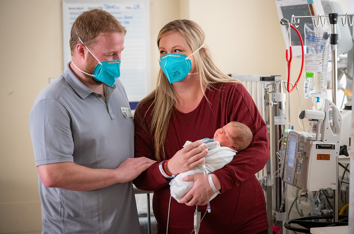
Michelle Johnson learned about her son’s diagnosis during a scheduled ultrasound study at the 20th week of pregnancy. Despite the fact that the family lived in Oregon, they decided to move to another state in order not to miss the chance of being involved in this experimental treatment. For Michelle, this was a rare opportunity to change her son’s future before he was born.

Today her son, Toby, is already four years old and his daily life is the best proof of the effectiveness of the study. He not only moves independently but runs, is active, and, as his mother says, is the real “boss” at home. The Johnsons are now sharing their experience with others as well, because they want Toby’s example to give hope to those parents who stand before a similar challenge.
Expert Assessments and Future Perspective
The medical community meets the results of the study with optimism, though with scientific caution. Gabriel Galea, an expert from University College London (UCL), emphasizes the project’s solid evidence base, though at the same time points to the necessity of long-term clinical monitoring. According to his explanation, years are needed for the risks of the development of neoplastic processes or the disturbance of kidney function to be finally excluded. While Professor David Chitayat of Mount Sinai Hospital believes that the regenerative potential of the method will increase even more in the future if a minimally invasive, fetoscopic surgery technique is added to it.
At this stage, the study is moving into the decisive 1/2a phase, which envisions long-term observation on 35 newborns. Scientists will follow their development until the age of 6 years, in order to evaluate in detail not only physical skills but also the functional state of internal organs with specialized neuro-motor scales (TIMP, Alberta, Peabody).
This project supported by the California Institute for Regenerative Medicine (CIRM) is a historical precedent, because it represents the first case of the prenatal use of stem cells in the treatment of a fetal neural tube defect.
Source: The Lancet
