


Amyotrophic Lateral Sclerosis (ALS) has, thus far, successfully resisted almost every drug deployed against it. While regulatory bodies periodically approve new treatments, the majority of clinical trials end in failure. Amidst these setbacks, the disease continues its steady destruction of the body. However, studying the regulatory mechanisms of the internal chemistry of motor neurons may explain why these specific cells become the direct targets of ALS, offering a therapeutic strategy that has never been fully utilized before.
What is ALS?
Amyotrophic Lateral Sclerosis, often referred to as Lou Gehrig’s disease, is a fatal condition. During this process, motor neurons—the nerve cells responsible for voluntary muscle movement—gradually die. As these cells are destroyed, patients progressively lose the ability to move, speak, eat, and breathe. For those facing this severe diagnosis, life expectancy generally ranges from three to five years.
ALS affects an average of 4 to 8 people out of every 100,000, resulting in approximately 30,000 fatalities annually. Despite decades of scientific inquiry, therapeutic options remain very limited. A case in point is the first authorized medication, Riluzole, which extends life by only 6 to 19 months. Given this context, the need for innovative treatment methods is clear.
The Selectivity of ALS
One of the most perplexing aspects of ALS is its selectivity. The destructive force of the disease is directed exclusively at motor neurons, while other elements of the nervous system remain largely intact. Although hypotheses exist regarding a unique biological “weak point” in motor neurons, the etiology of this phenomenon remains elusive to scientists.
A new study, conducted on the spinal cord tissue of deceased ALS patients and healthy donors, offers a new path toward solving this complex puzzle.
The Cellular Cleaning System
To better understand the essence of this discovery, one must first understand how a cell’s internal “waste processing plant” works. Our cells constantly produce proteins that, over time (like any other mechanism), become damaged, misfolded, or worn out. When this happens, the cell must break them down and recycle their components. This process is called autophagy (from the Greek for “self-eating”). While several types of autophagy exist, this study focuses specifically on a highly selective form known as Chaperone-Mediated Autophagy (CMA).
The working principle of CMA resembles a waste collection service. To identify, mark, and transport damaged or redundant proteins to lysosomes—the cell’s “cleaning stations”—the system utilizes a special “escort protein” called HSC70. In this process, the primary portal for entering the lysosome is the LAMP2A protein. LAMP2A can be viewed as a gatekeeper: the higher the concentration of this protein in the cell, the more active and efficient its CMA cleaning system becomes.
What the Researchers Discovered
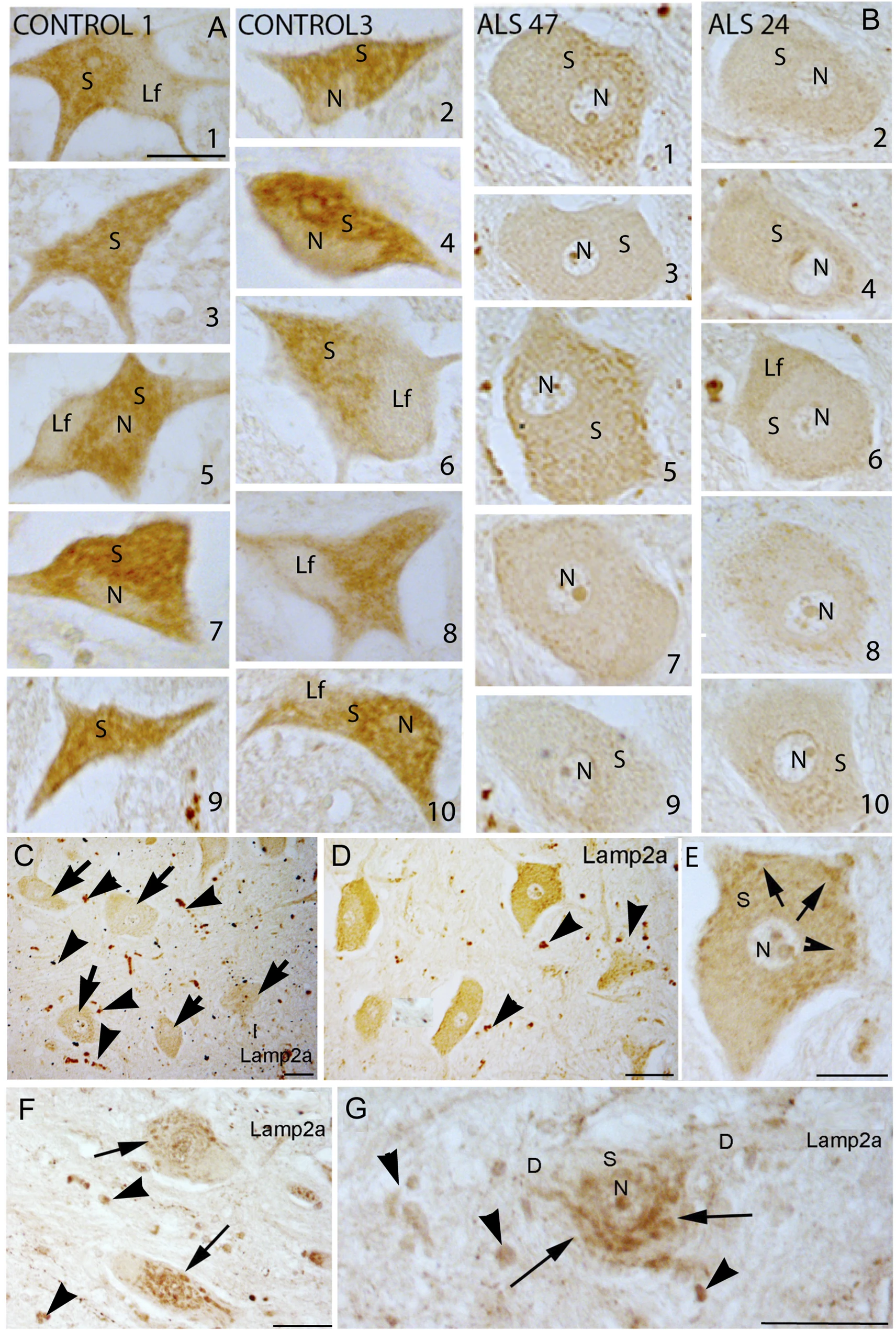
The research team compared the spinal cord tissues of ten ALS patients with those of six healthy individuals. The specialists paid particular attention to the concentration of the LAMP2A protein within motor neurons. The primary result of the study reinforced existing assumptions: compared to other nerve cells, healthy motor neurons contained significantly higher levels of LAMP2A. This suggests that the normal functioning of motor neurons is heavily dependent on the CMA process.
This phenomenon has a specific biological basis—motor neurons are among the largest and most metabolically active cells in the body. They operate continuously, possess a complex structure, and require a constant maintenance of protein balance.
The study’s second finding was even more striking and critical: in the motor neurons of people with ALS, LAMP2A levels dropped significantly. The “cleaning” mechanism stopped working precisely in the cells that need the system most. Importantly, this deficiency specifically affected only CMA. Scientists checked other forms of autophagy and found no impairments. Thus, the flaw was not in the general process of cellular waste recycling, but specifically within this particular pathway.
The Link to TDP-43
Flaws in the CMA system become even clearer when analyzing processes related to the TDP-43 protein. Under standard conditions, TDP-43 resides in the cell nucleus and actively participates in regulating genetic material. However, in nearly 95% of ALS cases—regardless of the cause of the disease—this protein leaves the nucleus and aggregates into harmful clumps within the cell’s cytoplasm. Such toxic formations are considered one of the fundamental hallmarks of the ALS pathological process.
The TDP-43 protein has a specific molecular marker that makes it a target for recycling by the CMA system. Scientists confirmed that in healthy cells, TDP-43 and LAMP2A coexist in the same space. This fact indicates that the CMA mechanism effectively destroyed harmful proteins before they could form aggregates. However, in the neurons of ALS patients where a LAMP2A deficiency was observed, TDP-43 accumulated in the exact pathological form that characterizes this disease.
What Protects a Specific Group of Neurons?
The most striking evidence from the study comes from a small cluster of neurons known as Onuf’s nucleus, located in the sacral part of the spinal cord. These neurons control the pelvic muscles and sphincters and exhibit remarkable resistance to ALS pathology. Because of this resilience, patients retain control over urinary and bowel functions until the advanced stages of the disease.
Naturally, the question arises: why do Onuf’s neurons maintain function while their neighboring cells are damaged? Scientists discovered that in ALS patients, the neurons of Onuf’s nucleus maintained high levels of LAMP2A. As a result, their CMA system continued to function without interruption. Thanks to this, TDP-43 remained in the nucleus in these cells, and toxic cytoplasmic clumps did not form. This picture exactly mirrors the biological state found in healthy tissue.
An Exceptional Case
The study identified one patient (coded as ALS40) whose motor neurons showed significantly higher levels of LAMP2A compared to other studied cases. Consequently, pathological changes related to TDP-43 were far less pronounced in their cells. Unlike other patients, this individual’s body cleared the toxic protein much more efficiently.
Notably, in the case of ALS40, the disease progressed much faster than in other patients. This phenomenon suggests a different underlying mechanism for the development of the disease. This case confirms once again that ALS is not a single, uniform pathology, but a broad spectrum fraught with individual variations.
What Happens in Glial Cells?
The study also evaluated the auxiliary structures of the nervous system, specifically glial cells. The researchers focused on a specific group of cells called astrocytes. In contrast to motor neurons, an increase in LAMP2A concentration was observed in these cells during ALS.
Scientists view this phenomenon as a response to inflammatory processes occurring in the surrounding tissues. In their view, this fact is not evidence of healthy CMA operation. It is clear that glial cells ramp up their waste processing mechanisms to cope with a crisis situation. However, such a reactive increase was still not enough to save the dying motor neurons. Furthermore, this circumstance likely masked the deficiency in motor neurons in earlier scientific works, as those studies did not strictly distinguish between cell types.
How Does This Discovery Change Things?
Scientists emphasize that this work is fundamental in nature. It describes a specific disease mechanism, but it is too early to say that enhancing CMA will definitely stop ALS. Nevertheless, the possibility of finding new avenues for treatment appears quite promising.
If the death of motor neurons in ALS is primarily driven by a breakdown in CMA, then stimulating this mechanism—specifically, artificially increasing the amount of LAMP2A—emerges as an entirely new therapeutic method. Certain experimental compounds that boost CMA activity are already being used to treat other neurodegenerative conditions, such as Parkinson’s and Alzheimer’s disease, where similar processes of harmful protein accumulation occur.
It is also vital to discover that in humans with ALS, macroautophagy—the other major mechanism of autophagy—functions unimpaired. This suggests that previous treatment attempts aimed at a general activation of autophagy processes were heading in the wrong direction. Consequently, the CMA system appears to be a much more precise and effective target for intervention.
Source: Acta Neuropathologica Communications
