


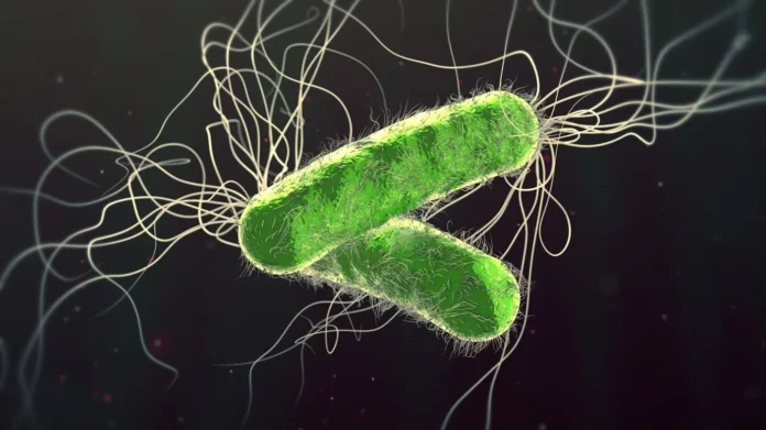
In the fight against bacterial infections, the human body often employs a strategy called “nutritional immunity.” During this process, the body attempts to strictly limit pathogens’ access to vital elements, primarily iron. However, some aggressive bacteria have developed unique mechanisms for survival and attack under these “starvation” conditions.
One such pathogen is Pseudomonas aeruginosa (blue-green pus bacillus). It is the primary causative agent of fatal pneumonia developed during cystic fibrosis and chronic obstructive pulmonary disease (COPD). For a long time, it remained unclear to scientists why this bacterium causes chronic, persistent infections in some cases, while in others, it leads to rapid and irreversible damage to lung tissue.
A study conducted to investigate this issue showed that the change in the bacterium’s behavior is driven by iron levels. Precisely this indicator determines the pathogen’s degree of toxicity.
The Role of Iron
Upon penetrating lung tissue, P. aeruginosa forms biofilms. These structures protect the pathogen from the effects of antibiotics. The bacterium’s virulence is determined by toxins such as pyocyanin, elastase, and exotoxin A.
In this process, iron represents an essential resource for bacterial growth. The host organism stores this element in specific proteins (e.g., transferrin), while the bacterium attempts to obtain it through siderophores. The study confirmed that healthy lungs keep pathogens in conditions of iron deficiency. In contrast, high concentrations of iron in diseased lungs facilitate the chronic course of the infection.
Analysis of strains isolated from patients and standard cultures showed that an excess of iron promotes an increase in bacterial mass and the formation of solid biofilms. However, the most unexpected result was revealed under conditions of iron deficiency. In such an environment, bacteria create weak protective barriers, but their virulence increases sharply.
The study showed that iron deficiency fully activates the bacterium’s virulence factors. When the concentration of iron decreases, the internal regulatory protein (Fur) ceases its inhibitory effect on toxin synthesis. As a result, the pathogen begins a massive release of toxins, leading to tissue degradation through the activation of elastase and protease. Simultaneously, the increased secretion of exotoxin A and pyocyanin results in the direct destruction of cells.
Experiments conducted on animals revealed a paradoxical picture. In an iron-rich environment, the bacterial population grows rapidly, but tissue damage is minimal. Conversely, under conditions of iron deficiency, the number of bacteria decreases, but acute pathological processes develop in the lungs. This state leads to alveolar collapse, severe edema, and large-scale hemorrhaging.
This research confirms that in the process of fighting infection, the number of bacteria alone does not have decisive importance. In this case, managing the pathogen’s behavior becomes a priority. Controlling iron homeostasis represents a new challenge for modern medicine. It is essential to inhibit bacterial growth through mechanisms that do not trigger their toxic activation.
წყარო: Frontiers in Microbiology
