


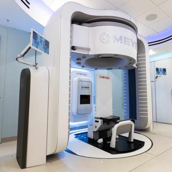
On April 7 of this year, a new department opened at the Stanford Medicine Cancer Center, where, for the first time in the world, pediatric and adult oncology patients will be able to receive ultra-compact proton therapy. This treatment method allows radiation to be directed with maximum precision directly at the tumor formation, with minimal damage to healthy tissues. Although this treatment approach was developed as early as the 1950s, the massive size of traditional proton therapy machines and the high cost of the procedure significantly limited the possibility of its widespread implementation.
The priority of using proton therapy increases in clinical cases where the neoplasm is located near critical anatomical structures—such as the brain, heart, spinal cord, or nerves involved in speech and swallowing. In these cases, protons have a significant advantage over traditional radiotherapy (X-ray/photon treatment), as the risk of collateral damage is minimized.
How Proton Therapy Works
Protons are tiny, positively charged particles located in the nucleus of an atom, whose physical properties (charge and mass) allow for precise control of their movement. By adjusting the energy, it is possible to aim protons so precisely that they stop inside the tumor; as a result, the “exit dose” is reduced to practically zero.
In contrast, photons used in conventional radiotherapy continue to move after passing through the tumor, which leads to the irradiation of healthy tissues located behind it. Strictly targeted protons help patients avoid complications caused by unintended radiation, specifically nerve damage, as well as organ dysfunction resulting from inflammatory processes and scarring. It is this ability to minimize damage to healthy tissues that makes proton therapy a priority for treating tumors of the brain, spine, head and neck area, lungs, liver, and prostate.
Minimizing side effects is especially important for children. “Because children are growing and developing, different parts of their bodies are more sensitive to even low doses of radiation,” noted Susan Hiniker, Associate Professor of Radiation Oncology at Stanford.
It is noteworthy that proton therapy is not a universal choice for all types of malignant tumors. Depending on the location and characteristics of the tumor, other radiotherapy methods may still remain optimal; however, proton therapy represents an important additional tool in planning individualized, case-based treatment.
Future Perspectives
While preparing to receive their first patients, the Stanford Medicine radiation oncology team is also working on further perfecting the technology and maximizing its accessibility.
Scientists plan to study the advantages of vertical (upright) irradiation compared to horizontal. According to existing data, in certain oncological pathologies, such as lung cancer, a seated position puts the organ in a more favorable state for safe radiation—the lung is more expanded in a vertical position, which reduces the risk of damaging healthy tissues. Additionally, the patient’s vertical position provides more flexibility for delivering radiation from various angles.
Currently, the Stanford Medicine Cancer Center is the only one in the world equipped with this latest proton therapy machinery. However, over the coming months and years, there are plans to implement this technology in other medical institutions and centers, which will ultimately make the treatment much more accessible.
In the U.S., approximately two-thirds of oncology patients require radiotherapy. “This means that any step forward in improving radiotherapy is a major gain for cancer therapy,” the scientists stated.
